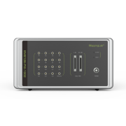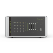
Electrical Mapping Systems
Applicable animals: Zebrafish, mice, rats, guinea pigs, rabbits, dogs, sheeps, pigs, monkeys.
Applicable tissues: cardiac slices and tissues, conductive tissues including sinoatrial node, atrioventricular node, Purkinje fibers, iPSC cells as well as in vivo and ex vivo heart, may also applicable to brain in vivo , brain slices, gastrointestinal tissues and uterus with customized system.
- High performance amplifiers and analog-digital converters (ADCs).
- Advanced acquisition and intuitive analysis software.
- Data transmission: USB or PXI fibre optic transmission.
- Various size and layout.
- Up to 16 additional channels.
- Multiple input/output (I/O) connectors.
- USB CCD camera.
- 12V DC power supply.
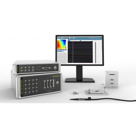
The Electrical Mapping Systems (EMS) are equipped with the new generation of Multi-Electrode Arrays (MEAs) and high-performance amplifiers. The advanced EMapping software facilitates fast recordings of field potentials from in vitro, in vivo, and ex vivo cardiac samples. The electrical activities and conductive information can be studied in great details at tissue level. The precision of data acquisition system makes it possible to accurately detect abnormalities on ion channels. It also can be grouped with other devices such as ventricular or coronary pressure probe, monophasic action potential system (MAP), myocardial tension, perfusion temperature and the optical mapping system. These features immensely empower cardiac electrophysiological researchers to understand better about mechanisms of cardiac arrhythmias. Our system is also an ideal candidate for efficiently screening new anti-arrhythmic drugs and testing drug toxicity.

