Irradiation - Applications of irradiation in the cell biology laboratory
Introduction
The development of specialized cell models for the study of antigens, antibodies, cell differentiation and cell death is an important activity in the cell biology laboratory. The creation of cells that: present the needed antigen to target (APC), produce antibodies to specific antigens (hybridomas), and feeder cells for stem cell research, require that, at some point, replication be slowed if not stopped.
Low level irradiation of biological tissues has many applications. In agriculture irradiation of seeds is one of the methods used to create new traits. In the cell biology lab, whether the lab is interested in stem cells, oncology or other applications, irradiation can be one of the tools to produce the biological material with the properties required.

Feeder cells
Cultures of feeder cells have been used for years to promote cell proliferation, particularly with low-density inocula. Basically, feeder cells consist in a layer of growth-arrested cells (unable to divide), which provides extracellular secretions (growth factors, adhesion molecules, and extracellular matrix) to help another cell to proliferate. Historically, either y-radiation (Cobalt source) or Mitomycin – C (MMC) has been used to arrest the cell growth, but the expense and availability of the Cobalt irradiators have been a hindrance for that method". The use of Feeder Cells is important in maintaining the health of the Stem Cell culture during development and differentiation.
APCs
Antigen-Presenting Cells (APCs) are heterogeneous immune cells involved in the cellular immune response. These cells mediate the immune response by processing and present antigens, surface molecules, which lymphocytes recognize. APGs include dendritic cells, macrophages, Langerhans cells and B cells.
Typically, the growth of APCs has been arrested by the use of Actinomycin-D, but irradiation has also been used?2
Hybridoma cells
A hybridoma cell is the fusion of two cell types for the production of specific antibodies. Hybridomas are produced by injecting the target antigen into a mouse, collecting antibody-producing cells from the mouse’s spleen, and fusing it with myeloma cells. The resulting cells are then treated to stop growth and screened for those that produce the desired antibodies. The immortalized cell line is then sed for the production of the antibodies. Irradiation can be used in many aspects of Hybridoma production including arresting of cell growth and preparation of feeder cells.
Apoptosis
Irradiation has also been employed in the study of cell death through the process of apoptosis. Apoptosis is cell death in response to stimuli (as compared to necrosis, cell death due to trauma). The irradiation is used as the stimuli to allow the study of the mechanism that cause apoptosis.3
X-ray verses isotopic source
The use of X-ray emitted by an electronic source versus X-ray emitted by radioisotopes (such as Cobalt and Cesium4) does not appear to change the result in biological assays.There is still of course the need to develop a proper irradiation protocol that takes intoaccount the variations of cell resistance to radiation, vessel holding the cells, cell thickness and media composition.
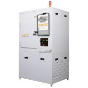
Benefits of adding benchtop irradiator to the laboratory
Asmall, bench top irradiator located in the cell biology laboratory offers the staff many advantages. There is improved efficiency due to reduced travel to a core irradiator. Since the bench top irradiators are electronic, generating X-ray, the security concerns with using radioisotope systems is removed. This makes not only use of the system easier, but also removes barriers to the purchase of the system.
Training and the requirement for background checks of the staff are also reduced, or eliminated. These systems do not require a special cooling system, and they can be plugged into a standard wall outlet. Since they are fully shielded (meeting the FDA requirements for cabinet systems), they do not have any costs associated to site preparation. They can easily be relocated within the lab space, or building (depending on local/state regulations).
There are additional safety benefits, since the use of chemicals such as Actinomycin D, Mitomycin — C (MMC) and Hypoxanthine-Aminopterin-Thymidine (HAT media) is reduced or eliminated. Since many of the core irradiators are located in the vivarium, the investigators reduce their exposure to the animal antigens.

References
1) Efficient feeder cells preparation system for large-scale preparation and application of induced pluripotent stem cells.
Pengdong Li, Shichao Wang, Lixiang Zhan, Xia He, Guangfan Chi, Shuang Ly, Ziran Xu, Yuhan Xia, Shuzhi Teng, Lisha Li & Yulin Li. Scientific Reports volume 7, Article number: 12266 (2017).
2) Peptide-b2-microglobulin-major histocompatibility complex expressing cells are potent antigen-presenting cells that can generate specific T cells.
Sonja Obermann, Susanne Petrykowska, Michael P. Manns, Firouzeh Korangy and Tim F. Greten. Immunology, 122, 90-97 (2007).
3) Regulation of Apoptosis aha Radiation Sensitization in Lung Cancer Cells via the Sirt1/NF-kB/Smac Pathway.
Kaihua Ji, Xiaohui Sun, Yang Liu, Liging Du, Yan Wang, Ningning He, Jinhan Wang, Chang Xu, and Qiang Liu. Cell Physiol Biochem 48:304-316 (2018).
4) Comparison of Cesium-137 and X-ray Irradiators by Using Bone Marrow Transplant Reconstitution in C57BL/6J Mice.
Brian W Gibson, Nathan C Boles, George P Souroullas, Alan J Herron, Joe K Fraley, Rebecca S Schwiebert, John J Sharp, and Margaret A Goodell.
Comparative Medicine Vol 65, No 3 Pages 165-172 (2015).