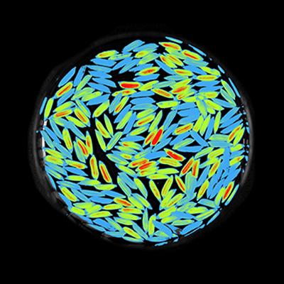
NEWTON 7.0 BIO for plant imaging
- Custom made f/0.70 lens: Specifically developed for macro imaging, the NEWTON 7.0 BIO proprietary optics offers you unrivalled sensitivity and sharpness.
- Multispectral imaging: Benefit from ultra-low noise imaging thanks to a dual camera amplifier architecture which ensures transmission above 90%.
- Narrow bandpass filters: With our technology based on very narrow band cutting, the time to get the image is drastically reduced and sensitivity is increased.
- Height adjustable plant stage: The stage can be tilted by 15° on the X/Y axis and is also motorized on the Z-axis. It can be easily controlled from the software interface.
Grow Your Plant Images
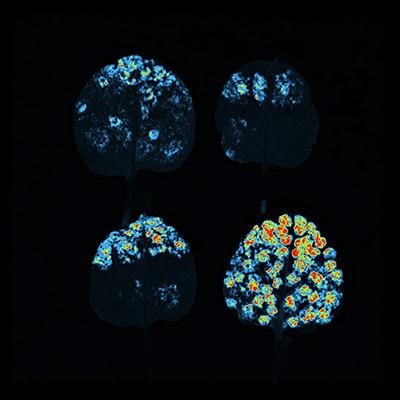
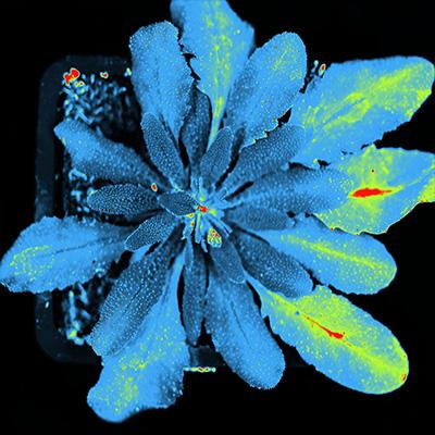
The NEWTON 7.0 BIO is a smart world-wide used optical imaging system dedicated to bioluminescence and fluorescence detection for plants and other in vivo / in vitro biological samples. Thanks to its unrivalled precision optics (ultra-low noise CCD camera and f/0.70 lens aperture) and its wide spectral range detection (400 – 900nm), the system allows you to cover all your applications. From visualizing infections in plants (like Arabidopsis thaliana) leaves and seedlings, to comparing plant virology, plant growth or plant stress tolerance, researchers will be able to benefit from an innovative and user-friendly interface to observe the lowest signal intensities in a live 3D sample reconstruction. In the end, the NEWTON 7.0 BIO is an instrument that is specifically made for absolute quantification and offers you the perfect mix between high sensitivity and deep analysis capabilities.
A wide range of applications
- Comparative plant virology
- Genetic regulation
- Infection monitoring
- Regulation of plant growth
- Stress tolerance
- Circadian rythms
Best camera performance
- Proprietary custom-made lens with the widest f/0.70 aperture in the industry
- 1” scientific grade CCD camera
- Deep CCD Cooling, -90°C via 4 Peltier
- Best Signal to Noise Ratio
- Ideal for faint luminescence applications
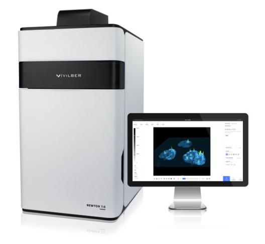
Incredible ease of use
- Unlimited number of users of the software
- Intuitive user interface
- One click to get the image
- Auto-exposure and automatic illumination control
- Adjustable stage
Minimum manipulation
- Up to 20x20cm FOV
- Motorized rotating stage on the Z-axis
- 15° inclination on the X/Y axis
- Control from the software
- Possibility to image whole plants, leaves and seedlings with unrivalled sensitivity and image resolution
This Is What Smart Means
Make publication-ready scientific images with simplicity for an unlimited number of users. Benefit from our 3D Dynamic Scan technology and observe the different signal intensities in a live 3D video reconstruction which will help you to detect signal concentration. The unique color imaging mode helps you acquire a quick snapshot of your plants with a true color representation, making the documentation faster! Our system’s protocol driven image acquisition is as quick as it is intuitive: adjust your exposure, save, print or quantify. Various imaging modes are available from automatic, manual, or time-lapse imaging program. Time series could be constructed from images acquired on different days. You can also monitor the growth of a plant overtime thanks to the daylight and nightlight simulation modes through the software. Finally, you choose the leaves you want to focus on with one click.
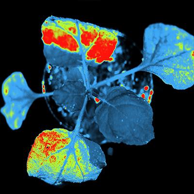
All The Applications You Need, In One Plant Imaging System
- The NEWTON 7.0 BIO allows you to perform all your delayed fluorescence, biophoton emission, luciferase / GFP expression and chlorophyll fluorescence applications. Visualize small plants, fruits, vegetables, leaves, cyanobacteria, green algae, seedlings, insects, blossoms, roots, mosses, lichens, fungi, cell and tissue cultures.
- Our system accommodates 8 excitation channels in the visible RGB and NIR spectrum (from 400 to 900nm). Signals can be overlaid so that several reporters can be visualized simultaneously. The very tight LED spectrum is additionally constrained with a very narrow excitation filter. This means less background in the images (ultralow noise imaging) of your sample and a higher signal to noise ratio to detect the weakest signals.
- A large number of dyes and stains can be used such as CFP, GFP, YFP, RFP, FITC, DAPI, Alexa Fluor® 680, 700, 750, Cy® 2, 3, 5, 5.5, DyeLight, IRDye® 680, 800CW.
