


Newton FT-30 Compact In Vivo Fluorescence and Bioluminescence Imaging System
Newton FT-30 Compact In Vivo Fluorescence and Bioluminescence Imaging System
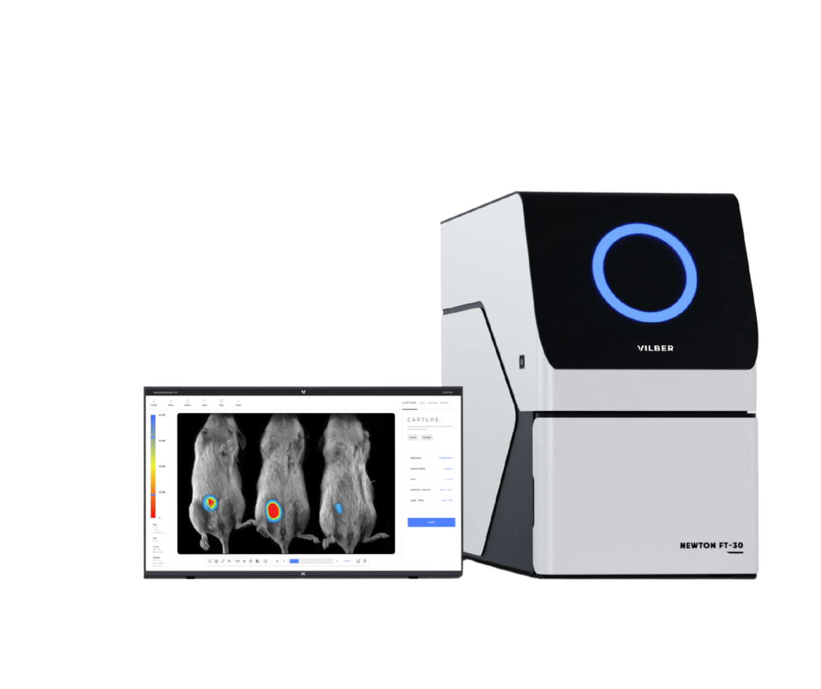
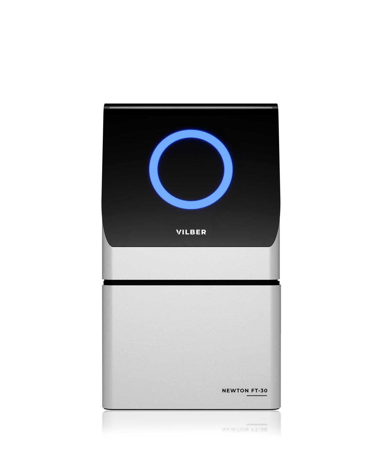
The Newton FT-30 is a compact, high-end optical imaging system for fluorescence and bioluminescence applications in vivo, ex vivo and in vitro. It combines an ultra-sensitive f/0.70 lens, a deeply cooled scientific-grade CCD camera and automatic analysis in a single benchtop unit, and it is designed to deliver absolute quantification of optical signals. Small animals, organs, tissues, Petri dishes and microplates can be imaged in the same system, making the Newton FT-30 a versatile tool for preclinical research laboratories.
Absolute Design
- Absolute calibration – imaging calibrated against traceable references from the National Institute of Standards and Technology (NIST) for precise quantification of specific biomolecules, such as proteins, with high accuracy, reproducibility and standardization
- Absolute quantification – unlike relative quantification, which only compares the amount of an analyte between samples, the system expresses results in absolute units such as radiance (ph/s/cm²/sr), so data can be compared and integrated across studies and laboratories
- Absolute efficiency – a wide door opening makes it easy to place and arrange samples; once inside, the system automatically sets the camera position according to the nature and size of the sample
- Absolute versatility – a customizable and upgradeable configuration with a choice of 8 Spectra LED excitation sources from 400 nm to 800 nm for flexible fluorescence imaging
- Absolute durability – a robust aluminium and stainless-steel construction, optimized for a compact footprint without compromising high-resolution imaging
Performance and Detection
- Detection at the limits – the ultra-sensitive f/0.70 lens is optimized for faint-light conditions down to the picogram level, and absolute cooling to -90°C provides a high signal-to-noise ratio for detection as low as a single cell
- Premium optics – the very low f-number means a wide aperture that lets more light reach the sensor, for faster imaging and higher sensitivity; the deeply cooled scientific CCD camera minimizes background noise
- Multispectral imaging – images captured at multiple wavelengths extract comprehensive information from a sample and improve the accuracy, versatility and efficiency of the imaging process
- Immediate results – ultra-sensitive detection allows shorter excitation exposure and reduced acquisition times; protocol-driven acquisition is quick and intuitive: adjust the exposure, then save, print or quantify
Absolute Analysis with KUANT Software
The system transforms light into quantitative data on user command, while the user keeps control over the analysis to preserve data integrity. Results are displayed as calculated values overlaid on the acquired image, making them easy to communicate. By combining multispectral imaging with spectral unmixing, the KUANT software addresses autofluorescence challenges, and its N.M.F. (non-negative matrix factorization) algorithm improves detection accuracy. The license-free software supports full GLP compliance and includes absolute quantification, 3D Dynamic Scan, autofluorescence correction and kinetic imaging.
Luciferase Expression and Fluorescent Reporters
The Newton FT-30 visualizes a wide range of bioluminescent reporters, such as firefly luciferase, as well as fluorescent molecular reagents in the visible and NIR-I windows. Chemiluminescence imaging is also supported.
Animal Handling Features
- 3-mouse breather for imaging up to three anesthetized mice at a time
- Macro mode with a 6 × 6 cm field of view and a black anti-reflective surface for detailed imaging of organs and small samples
- Heating stage (Off / 37°C) to maintain animal body temperature during imaging
- Optional anesthesia and induction box
Applications of Newton FT-30
- Tumor and infection monitoring
- Cell migration tracking
- Targeting of molecules and nanoparticles
- Visualization of vasculature and microcirculation
- Biodistribution and pharmacokinetics studies
- Luciferase expression and fluorescent reporter imaging in in vivo, ex vivo and in vitro samples
For research use only.
- Imaging modes: bioluminescence, chemiluminescence, multispectral fluorescence
- Sample types: in vivo (small animals), ex vivo, in vitro
- Lens: custom-made, motorized, f/0.70
- Camera: scientific-grade 16-bit CCD, Grade 0 (zero defect)
- Cooling: -90°C with 4 Peltier stages
- Image resolution: 10 megapixels
- Native resolution: 2160 × 2160 pixels
- Fluorescence channels: 8 (excitation / emission): 440/500 nm, 480/550 nm, 540/600 nm, 580/650 nm, 640/700 nm, 680/750 nm, 740/800 nm, 780/850 nm
- Excitation sources: Spectra LED, 400–800 nm
- Filter wheel: motorized, 10 positions
- Camera positioning: Z-axis motorized camera with automatic zoom and sample position recognition
- Macro mode: 6 × 6 cm field of view, black anti-reflective surface
- Animal handling: 3-mouse breather, heating stage (Off / 37°C), optional anesthesia and induction box
- Hardware: intelligent darkroom concept, fully automatic system, software-controlled lighting with automatic visible lighting adjustment
- Construction: aluminium and stainless steel
- Software: license-free, full GLP compliance; absolute quantification, 3D Dynamic Scan, autofluorescence correction, kinetic imaging, spectral unmixing (N.M.F. algorithm)
- Calibration: NIST-traceable