Imaging - 3D capture vs. caliper for tumor measurements in laboratory animals
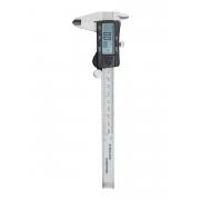
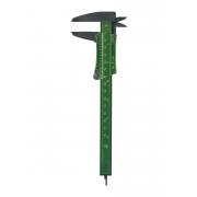
Caliper measurement of lab animal tumors
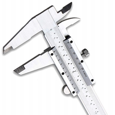
By using a caliper, one will measure width and length of the tumor and then calculate the volume using a standard formula, such as π/6 x W x W x L or 1/2 x W x W x L thus assuming a standard and constant shape of the tumor.

Using a caliper, growth of the tumor in height is not measured as only width and length changes will affect the calculated volume. This results in the loss of information about its real growth.
With constant width and length, the measured volume does not change with the tumor shape. The height and topography of the tumor is not taken into account in a caliper measurement.

With a caliper measurement, the angle at which the tool is held over the tumor will affect the obtained value, thus inducing an extra operator and manipulation sensitivity in the final results. This effect is illustrated in the included video.
A caliper only digitizes the length and width of the tumor. Further geometrical or visual data are not stored simultaneously.
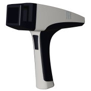
Direct 3D capture by Peira scanner

A direct 3D volume capture instrument, such as the TM900, will make a stereographic image of the tumor and reconstruct the tumor topography and shape. A direct and real 3D volume of the xenograft will then be calculated.

Monitoring tumor volume through a direct 3D measurement will include changes in all three directions, height, width and length thus resulting in more accurate realistic data.
With constant width and length, the measured volume changes with the shape of the tumor. The obtained data include height and topography changes.

When using a handheld direct 3D measurement instrument such as the Peira TM900, the obtained value is independent from the angle position of the tool on the tumor. How the device is positioned over the tumor will not influence the result.
The TM900 also stores an optical image in the measurement data base. These together with the tumor shape and all obtained data can easily be traced and consulted afterwards.
Measurement time
Both methods are comparable when it concerns the time it takes to do a measurement. For the TM900 one has to push the acquisition button and almost immediately the images are stored and values calculated. The manipulation of the handheld tool is at least as easy as holding a measurement caliper. For both one has to have the ease of hand and get used to manipulation of the instrument. Timing wise both methods perform the same.
Measurement method and operator dependency
When using calipers there is dependency in the measured data of the positioning by the operator of the tool on the xenograft. A direct 3d measurement tool such as the TM900 does not induce such method sensitivity but cannot exclude operator dependency of the obtained values. Both methods cannot avoid variability introduced by the manipulation of the animal. Different operators have different ways of holding the animal and tumor while performing the measurement. Even the same operator, with different trials or at different moments in time does not always hold the animal in exactly the same way. The tumor can be more and sometimes less visible or popped up from under the skin. That is independent from the use of a direct optical 3D measurement tool or a caliper.