Oxygenation - Monitoring oxygen in the eye
Oxygen, an essential element for life, plays a pivotal role in various biological processes such as cellular respiration, metabolism, and immune response. In the human body, oxygen is transported from the lungs to other tissues via the bloodstream, including the eyes, where it is crucial for the health and functionality of the cornea, retina, and other ocular tissues. A lack of sufficient oxygen supply to these tissues can result in eye disorders such as corneal edema, retinal diseases, and glaucoma. Therefore, understanding oxygen levels in the eye is pivotal for ophthalmology research.
In this article, we will discuss how Oxford Optronix has contributed to ophthalmology research, particularly in the area of oxygen levels and their effects on the eye. We will also summarize selected peer-reviewed publications that utilize our specialized systems for a better understanding of oxygen in an ophthalmology research setting.
Accurate pO2 measurements of the eye
Oxygen is vital for maintaining the health and function of the cornea and retina in the eye. The cornea, being avascular, relies on the surrounding atmosphere and tear film for its oxygen supply. Disruptions to this supply can result in corneal hypoxia, causing symptoms such as blurred vision and severe eye pain. The retina, one of the highest oxygen-consuming tissues in the body, requires an adequate oxygen supply for photoreceptor function and survival. Oxygen deprivation, or hypoxia, can lead to retinal diseases such as diabetic retinopathy and age-related macular degeneration.
Research on oxygen in the eye primarily focuses on understanding oxygen metabolism in ocular tissues and its impact on various eye conditions or diseases. This involves measuring the oxygen levels in different parts of the eye, such as the cornea, anterior chamber, vitreous humor, and retina. Taking these measurements in a delicate structure like the eye presents unique challenges. Various techniques have been developed for this purpose, and we have been involved in several studies that have used our OxyLite sensors for precise and direct oxygen measurements in these tissues. Our fine, bare-fiber sensors offer a minimally invasive way to measure different tissues and structures directly within the eye with pinpoint accuracy.
Modulation of oxygen in ocular cell models
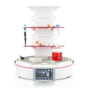
In addition to direct tissue oxygen measurements within eye structures, researchers also use cell models to understand the underlying mechanisms of cell function. Cells can be cultured in vitro and subjected to hypoxic conditions to study the effects of oxygen deprivation on cell function and survival. The HypoxyLab™ workstation provides a controlled environment for cell growth, regulating temperature, humidity, CO2, and most importantly, oxygen levels.
It's worth noting that while in vivo and clinical eye oxygen studies typically express their oxygen measurements in partial pressures (mmHg or kPa), most scientists conducting cellular studies in hypoxic/physoxic workstations refer to oxygen concentration in relative units of percent. The HypoxyLab bridges this gap by relying on absolute measurements of oxygen in mmHg to control its internal environment. This is not only scientifically more rigorous but also allows scientists to recreate conditions that replicate those experienced physiologically.
Preclinical publications
Park YH. et al. 2010 (British Journal of Ophthalmology)
Comparison of two probe designs for determining intraocular oxygen distribution
Despite the study's age, it remains an excellent resource for understanding the capabilities of our OxyLite sensors when comparing it to a polarographic electrode in ocular studies. The study cites the predecessor of OxyLite (the “OxyLab™ pO2” system) and found that the optode sensors is employs provide more accurate data in standard physiological ranges, compensate temperature in the eye more effectively, and generate pO2 data in the vitreous faster than the electrode. This study serves as compelling proof of concept for using fiber-optic sensors for direct pO2 measurements in the eye.
Fayyaz M. et al. 2021 (Applied Nano Materials)
Dextran-Based Oxygen Nanobubbles for Treating Inner Retinal Hypoxia
This article explores the use of oxygen nanobubbles in rat eyes to counteract the effects of central retinal artery occlusion (CRAO), a potentially blinding ischemic event.

The researchers utilize OxyLite sensors and demonstrate that the proposed treatment increased the oxygen availability in the eye at various depths, suggesting potential future emergency treatment for CRAO or similar ischemic conditions.
Williamson BK. et al. 2018 (Translational Vision Science & Technology)
The Effects of Glaucoma Drainage Devices on Oxygen Tension, Glycolytic Metabolites, and Metabolomics Profile of Aqueous Humor in the Rabbit
As glaucoma drainage devices (GDD) can lead to corneal decompensation, in this study investigators wanted to better understand this phenomenon and the impact GDDs had on several important eye related factors, including oxygen tension.
The study finds that oxygen levels in rabbit eyes were significantly lower two months post-surgery, indicating a need for further research to understand the correlation between GDD implantation and corneal decompensation.
Murali K. et al. 2016 (PLOS ONE)
Spatial Variations in Vitreous Oxygen Consumption
This research looked at spatial variation of vitreous oxygen consumption in ex vivo porcine eyes. Using the setup below the authors mapped the oxygen tension at 1mm steps away from the oxygen source to understand how much oxygen was being consumed in 2 separate regions of the eye.

The study revealed that vitreous oxygen consumption is higher in the posterior vitreous compared to the mid-vitreous.
Ye X. et al. 2024 (Molecular Therapy Nucleic Acids)
A novel function and mechanism of ischemia-induced retinal astrocyte-derived exosomes for RGC apoptosis of ischemic retinopathy
This study was the first eye-based cellular paper to employ and cite our HypoxyLab workstation. The researchers were able to simulate retinal ischemia within their cell models, leading to the discovery that hypoxia-stimulated astrocytes secreted exosomes carrying miR-329-5p, which appears to protect certain cells from ischemia-related apoptosis.

This finding offers promising future applications for treating retinal ischemia-related ocular disorders.
Clinical publications with OxyLite monitor
Please note: The OxyLite (or its predecessor system, the OxyLab pO2) do not possess FDA or clinical approvals for use in the human eye and are intended for preclinical use only. Any clinical use of these devices was approved independently by the investigators' respective internal review boards.
Huang AJW. et al. 2015 (Investigative Ophthalmology & Visual Science)
Impact of Corneal Endothelial Dysfunctions on Intraocular Oxygen Levels in Human Eyes
Here the authors sought to understand the effects of corneal endothelial dysfunctions on oxidative stress in the anterior chamber (AC) of human eyes.

The study found that eyes with endothelial dysfunctions had increased levels of pO2 in the anterior chamber compared to the control, suggesting that reduced oxygen consumption in dysfunctional corneal endothelial cells may increase oxidative stress in the anterior chamber.
Lange AK. et al. 2011 (American Journal of Ophthalmology)
Intraocular Oxygen Distribution in Advanced Proliferative Diabetic Retinopathy
Here this group explored the variances in intraocular oxygen distribution in patients with advanced proliferative diabetic retinopathy. The findings revealed a noteworthy decrease in oxygen levels in the anterior and mid-vitreous regions of the diabetic group. Conversely, there was an increase in oxygen concentration at the posterior pole.

The researchers attribute this to the direct correlation between the posterior pole in diabetic individuals and the elevated vitreous VEGF concentration. This could potentially trigger the formation of neovascular complexes in the retina, thereby enhancing oxygen supply to the area.
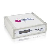
The OxyLite™ oxygen monitor
OxyLite™ - The gold standard tissue pO2 and in vitro dissolved oxygen monitoring platform, trusted by a worldwide user-base in research institutes, universities, hospitals, pharma, and CROs.