

Push-Button Image Analysis Software for Hermes microscope
Athena is a rich and simple application-based image analysis software for easy and quick evaluation of cell biology experimental results. Athena performs common and advanced cell biology applications at the push-of-a-button.
WiScan Athena is a software tool for application-derived analysis and visualization of image-based experiments. The software incorporates embedded analysis algorithms, statistical evaluation tools and sub-population analysis tools, based on advanced cell quantification tools for measuring fluorescence intensity and morphological features. It consists of a library of pre-built analysis modules applied to data sets of both labeled and non-labeled cells, thus Athena is capable of analyzing images acquired in bright field and fluorescence illumination microscopy.
Easy to use, push-button operation
All level users can be easily trained and use this platform simply and independently. Athena offers a bank of ready-made applications which are loaded by the push of a button and allow the user to start analysis within a few minutes, after performing mild parameter calibrations. The UI and UX are optimized for analyzing image-based cell biology experiments. New comers to high content image analysis can get productive right away using Athena’s pre-configured templates for the most common cell biology applications.
Analysis can be performed on a cell-by-cell basis and at population level.
Individual cells can be measured & quantified both for single cell analysis and for subpopulation analysis. Athena contains a population tool which in similarity with cytometry experiments, can be used to define sub-populations of cells in each well and quantify properties of this sub-population based on cellular phenotypes.
Image acquisition and image analysis can be done simultaneously, independently & at separate locations.
View and analyze data from any computer that has an installation of the Athena software, freeing up the Hermes imager for others to use. Experiment time is significantly reduced as images acquired in Hermes system are being uploaded in real-time to a shared storage, from which Athena software extracts data for analysis, in parallel to the image acquisition process. Experiment metadata, such as filters, objectives, plate type, fields & coverage, time points, z-stack or exposure, are stored in the database for future reference.
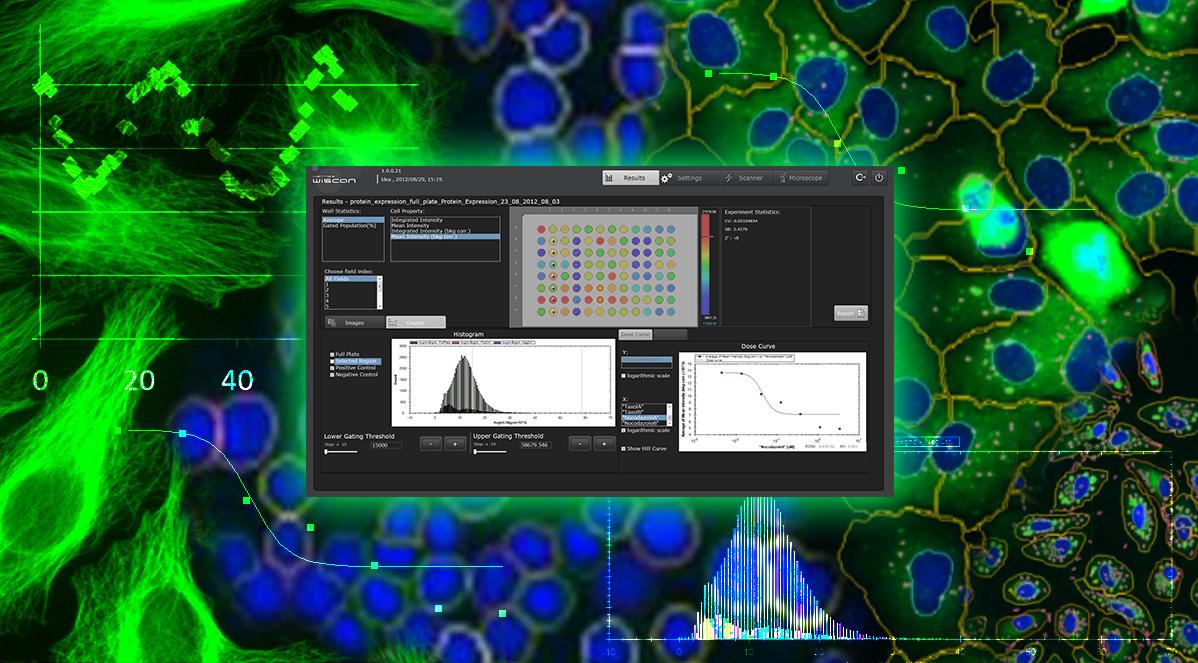
Straightforward, simple visual results display.
Athena software offers various visualization tools to display the analysis results. At the end of each analysis process, the user can quickly & easily create a summary report summarizing the analysis parameters and presenting the results. Raw data can be easily exported as an excel file to external sources if necessary.
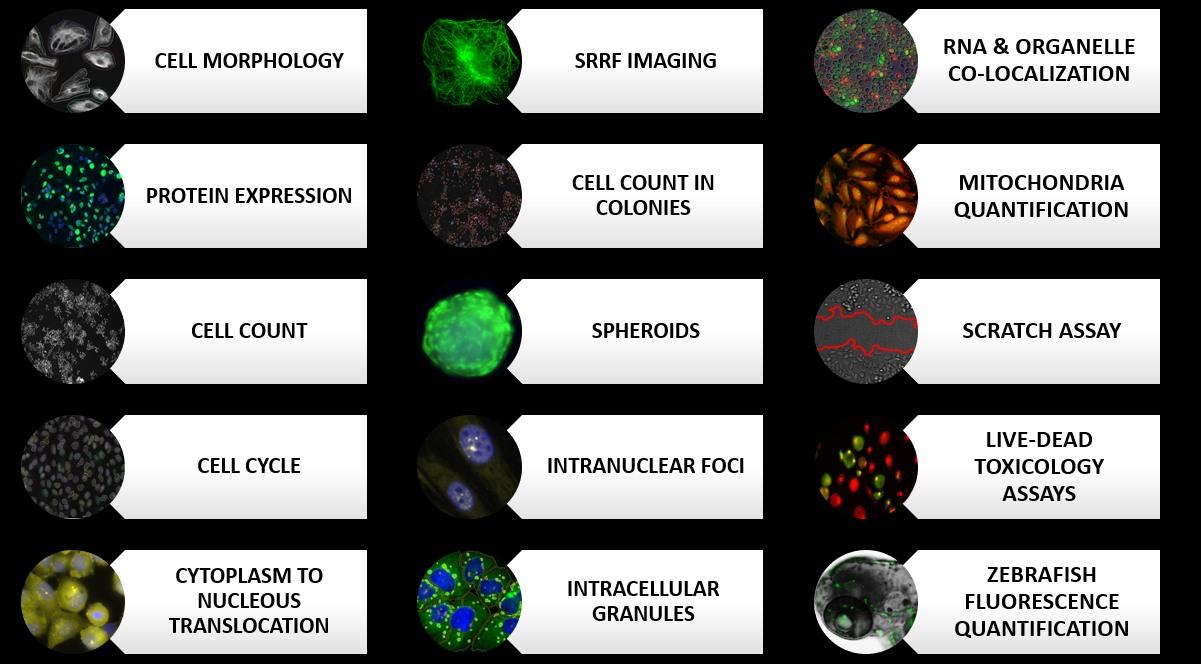
Athena software includes all the following cell biology assays as read-made applications:
Cell count:
Autophagy; Apoptosis; Cytotoxicity; Cell Viability; Cellular proliferation; Growth rate; Viral plaque assay
Cell morphology
Sub-cellular features quantification:Nuclei; Actin; Microtubules; Golgi; Vesicles; Focal adhesions; Mitochondria
Protein expression
Cell Health assessment; Protein degradation; Protein Localization; Protein translocation; Intracellular distribution analysis; Autophagy (t1/2) assays; Ubiquitination; Protein Aggregation & Accumulation; Transfection efficiency; Gene therapy; Pharmacology & pharmaco-kinetics
Cell cycle
Proliferation assays; Cell cycle arrest detection; Cytotoxicity; Cell Viability
Translocation
Nuclear signaling assays; Innate immune response; Intra-organelle localization; Intracellular distribution analysis
Athena software offer also specialized imaging applications:
Intracellular granules
Protein aggregation; Vesicle trafficking; Puncta analysis; Endo/exocytosis; Receptor or protein clustering
Cell count in colonies
Bacterial colony cell count; Small, high density cellular colonies e.g. Embryonic stem cells
Intranuclear foci
Intranuclear protein aggregation; Nucleolar detection; Transcription factor detection; Chromatin detection
Quantitative cytometry
Cytotoxicity; Autophagy; Apoptosis; Cellular fusion; Multi-plasmid transfection; Live/Dead measurement; Bacterial viability; Multi-color foci/puncta
Live-dead toxicology
Cytotoxicity; Toxicology; Cell viability; Pharmacology & pharmacokinetics
Spheroid Morphology: Drug dose response curves; Cytotoxcitity; Toxicology; Growth curves; Pharmacology & pharmacokinetics
Colony detection
Clonogenic assay; Radiotherapy assay; Dose response curve; Cell viability & proliferation; Pharmacology & pharmacokinetics
Yeast quantification
Cell replication & proliferation; Genetic profiling; RNA colocalization with intracellular organelle in yeast
Fiber detection
Cytoskeletal rearrangement
Confluency
Cell viability & proliferation; Radiotherapy assay; Growth rates; Population pharmacology & pharmacokinetics
Mitochondria quantification
Cellular metabolism; Cell health and viability; Mitochondrial dynamics: Number; Size; Distribution
Colocalization
Protein-protein interactions; Intracellular transport; Endo/exocytosis; Subcellular drug sequestration
Cell count in brightfield
Cell viability & proliferation; Radiotherapy assay; Growth rates; Pharmacology & pharmacokinetics
Zebrafish
Bacterial infection; Metastasis growth assay
C. elegans
Cell type identification
Scratch assay
Wound healing scratch assay
More information you will find on IDEA Bio-Medical website.