


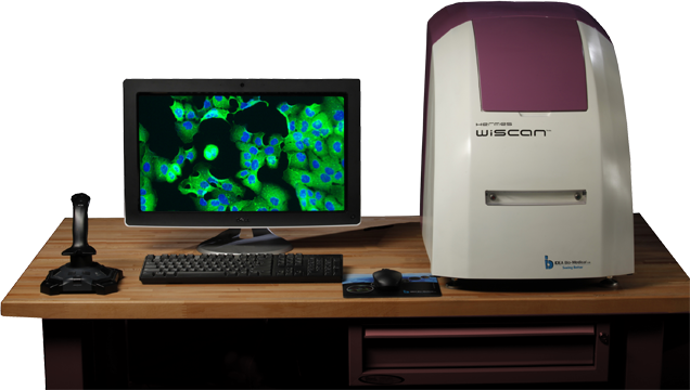


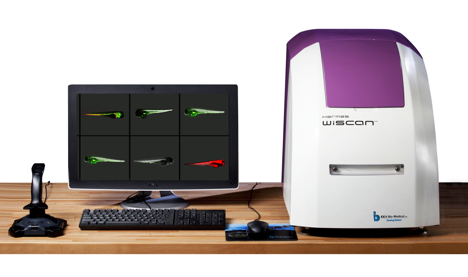
High Content Imaging System - WiScan Hermes
WiScan Hermes provides easy publication generations with quality images at high throughput speeds. Hermes microscope models have various configurations for use in the pharmaceutical industry and life science research.
Wide range of Hermes Industrial imaging system applications:
- Spheroid & organoid imaging
- Cell viability, cytotoxicity, autophagy, apoptosis
- Dose response assays
- Characterizing disease mechanisms, drug mechanisms
- Gene therapy assays
- Lipid accumulation assessment for drug effect studies
- Assess protein-protein interactions in living cells with Fluorescence Resonance Energy Transfer (FRET)
- FRAP for protein in-vitro
- MDR - characterize Multi Drug Resistance in cancer cells
Applications of Hermes Lab Partner and Real Time Observer models:
- Cell viability, cytotoxicity, apoptosis, autophagy
- Microbial assays - bacteria counting, bacteria viability & vitality
- Proliferation assays
- Endocytosis, phagocytosis
- Cell adherence, cytoskeleton & adhesion studies
- Proximity assays, Golgi cells tracking
- Protein localization & translocation, protein degradation assays (t1/2) / ubiquitination
- Protein aggregation and accumulation assay, dose response
- "In-Cell Western" - gross assessments of native protein expression / phosphorylation
- Transfection efficiency assay
- Subcellular distribution analysis
Main features of Hermes imaging system
Reliable
WiScan Hermes, IDEA Bio-Medical’s automated imaging system for high content screening (HCS) provides the unique combination of the two contradicting primary functions of automated microscopy: Image quality and acquisition speed.
Robust
WiScan® Hermes’ mechanisms are based on patents, creatively designed to meet heavy duty operation demands (24/7) with full process robustness.
Flexible
WiScan® Hermes is a cost-effective system that is both sophisticated and flexible, offering 7 fluorescence colors, bright field option, and a large range of air objectives. The system can accommodate a variety of multi-well plates and sample formats (slides, dishes…) and offers environmental control for live cell assays.
Intuitive
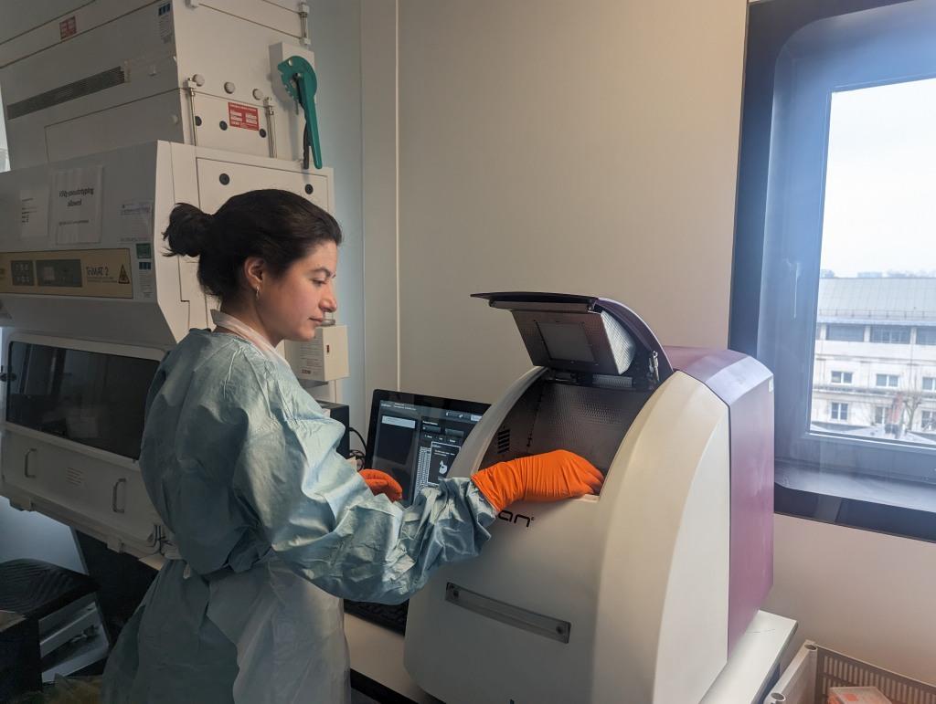
It doesn’t matter if you are a beginner or an experienced microscopist, Hermes easily allows you to look deeper into your samples. The system is intuitively operated. Its built-in applications are extremely easy to use, and are operated at the push-of-a-button.
Flexible plate formats & sample holders for WiScan Hermes system
- Supports full-area screening of 6-1536 well plates
- Holders for additional sample formats are available:
- Slide holder
- Chamber holder
- Dish (3cm) holder
- Histology slides, Spotted Array
- Any SBS-standard format can be easily introduced to the Hermes system using a simple definition screen, for maximum flexibility
Objectives selection for Hermes imaging system
Hermes system incorporates an automated objective exchanger which holds up to 3 objectives at a time. Hermes supports air objectives and can optionally support a unique immersion media dispensing to allow oil/water immersion objectives.
Objectives with air as immersion medium
- 2X, NA: 0.08, 3.0 µm/pixel, working distance: 6200 mm
- 4X, NA: 0.16, 1.5 µm/pixel, working distance: 13000 mm
- 10X, NA: 0.40, 0.6 µm/pixel, working distance: 3100 mm
- 20X, NA: 0.45, 0.3 µm/pixel, working distance: 6600-7800 mm
- 20X, NA: 0.80, 0.3 µm/pixel, working distance: 600 mm
- 40X, NA: 0.60, 0.15 µm/pixel, working distance: 2700-4000 mm
- 40X, NA: 0.75, 0.15 µm/pixel, working distance: 510 mm
- 40X, NA: 0.90, 0.15 µm/pixel, working distance: 200 mm
- 60X, NA: 0.95, 0.1 µm/pixel, working distance: 200 mm
Objective with water as immersion medium
- 20X, NA: 0.7, 0.3 µm/pixel, working distance: 350 mm
- 40X, NA: 1.15, 0.15 µm/pixel, working distance: 250 mm
- 60X, NA: 1.2, 0.1 µm/pixel, working distance: 280 mm
Objectives with oil as immersion medium
- 40X, NA: 1.4, 0.15µm/pixel, working distance: 130 mm
- 60X, NA: 1.42, 0.1µm/pixel, working distance: 150 mm
- 100X, NA: 1.45, 0.065µm/pixel, working distance: 130 mm
Hermes Real-time Observer data sheet
3D reader: EPI-fluorescence inverted optics mounted on XYZ (patented) linear scanner
- Auto Focus: Patented ultra-fast laser-based Auto Focus with 100nm resolution
- XY motion: Accurate positioning with 200nm repeatability
- Illumination sources: Hermes Real-time observer includes 4 fluorescence channels DAPI,,GFP,,RFP,CY5.
- Up to 7 optional LED sources are available in other Hermes models (DAPI,CFP,GFP,YFP,RFP,mCherry,CY5).
- Transmission: White LED source
- Optical Filters: 2 emission filters and compatible dichroic filters (automatically exchanged)
- Objectives (Air): Choice of air objectives in the range: 2X to 60X, high NA.
- Oil/ Water immersion objectives: Option to add automated immersion media to allow use of immersion objectives (oil/water) – optional hardware upgrade.
- Camera: High sensitivity CMOS camera with 5.1MPixel resolution
- Sample format: Supports full-area screening of 6-1536 well plates Supports slides, microarrays, 35 mm dish formats. U-shaped bottom plats are optional. Flexible and simple interface for introducing new sample formats to the microscope.
- IT: PC with Windows® operating system and touch screen. Joystick for microscope navigation.
- Enclosure: Allows operation in fully lit areas
- Desktop standalone platform: 47 W x 72 D x 57 H (cm), 18.5 W x 28.5 D x 22.4 H (inches) With plate cover closed.
- Certification: CE, UL
- Live cell imaging: Allow long time lapse experiments. Sample does not move during most of the scanning process.
- Live cell conditions in Hermes WiScan microscope
- Temperature control: Ranges from ambient+5°C to 40°C ±0.5°C
- CO2 level control: enables setting of 0.02 L/min- 0.13 L/min mixed with air for desired CO2 percentage. (external accessory). CO2 Range: 0-15%. Set Point Resolution: 1%
- Humidity
- Object mapping for rare events detection
- First scan: rapid scan of entire plate with low magnification, for efficient region of interest object mapping.
- Second scan: re-visitng solely detected objects with high magnification
- Analysis tools: WiSoft® based image processing tools for object definition and detection in the first low magnification scan and further tools to analyze the detected objects with high magnification
- Objectives: Automatic objective exchanger, accommodates of up to 3 objectives at a time
- Throughput: Enhances the throughput of high magnification scanning of rare objects
- Statistical tools: Provides automatic decision resulting in efficient screening for minimum required objects in a well
- Combination of HCS and HTS: Ultra-fast High Content Screening unit
- Image acquisition speed >10 images per second (depends on experiment conditions)
- Throughput / Acquisition speed: A full 96-well plate screening using 10X magnification with a single field per well, four fluorescence colors, 50ms exposure time per channel, runs in ~2 minutes. With some applications, this will include data processing and analysis results simultaneously with image acquisition.
- Automation Remote accessibility: Protocols ( applications and screening parameters ) execution and management
- External loader compatibility to 3rd-party robotics (Robot/manipulator)
- Communication channel for control by external equipment
- Loading door with automatic opening/closing synchronized with external equipment
- External loader(Robot/manipulator) integration: Full integration supported
| Product name | |
|---|---|
| Hermes Lab Partner Hermes imaging system, 2 fluorescence channels , cytotoxicity sampled assay; entry level | |
| Hermes Real Time Observer Hermes imaging system, 4 fluorescence channels, proliferation & translocation sampled assay, live cell basic | |
| Hermes Real Time Observer Pro Hermes imaging system, 4 fluorescence channels, protein accumulation & degradation, granularity sampled assay, live cell High Throughput screening | |
| Industrial HT - Hermes Drug Discoverer Hermes imaging system, 4 fluorescence channels, spheroid & organoid imaging sampled assay, automation interface to robotic arms | |
| Industrial HT 7 clrs - Hermes Drug Discoverer Pro Hermes imaging system, 7 fluorescence channels, multiplexed drug studies sampled assay, automation interface to robotic arms | |
| Industrial Live Cell HT 7 clrs - Hermes Supreme Vision Hermes imaging system, 7 fluorescence channels, imaging cytometry sampled assay, live cell experiments |