

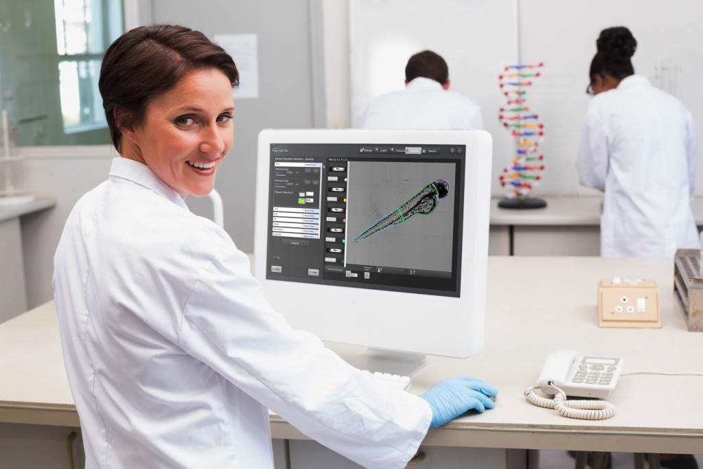


Zebrafish Image Analysis Software for Hermes screening system
Athena is unique dedicated analysis software for automated analysis of Zebrafish microscopy images. It offers simple and quick quantification of fluorescence, measurement of morphological changes & other phenotypic features in Zebrafish larvae in a high throughput format.
Athena Zebrafish is suited for a broad range of researchers and accepts multiple image format types output from nearly all microscope manufacturers.
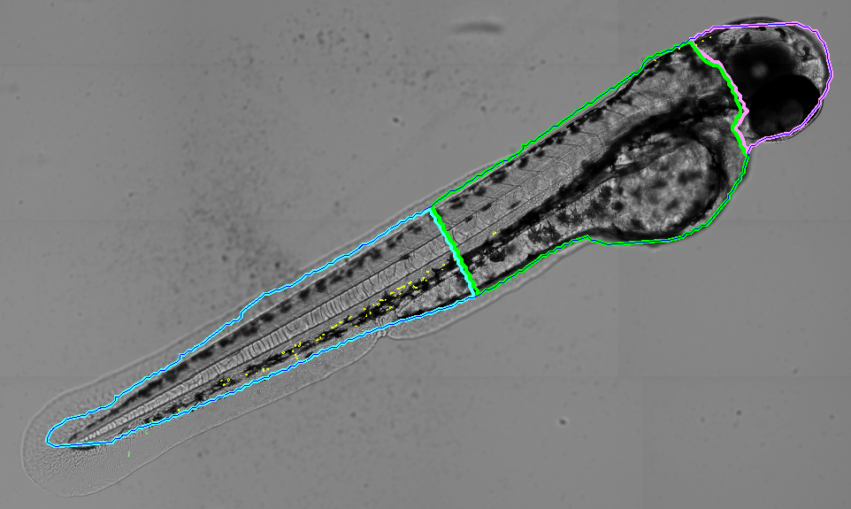
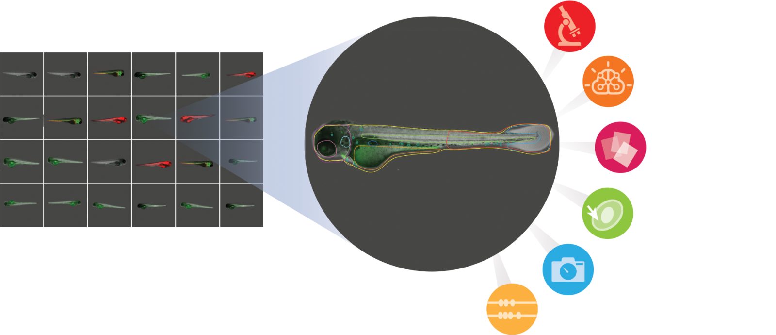
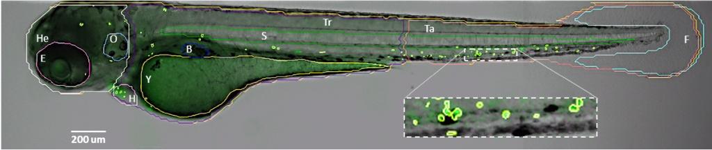
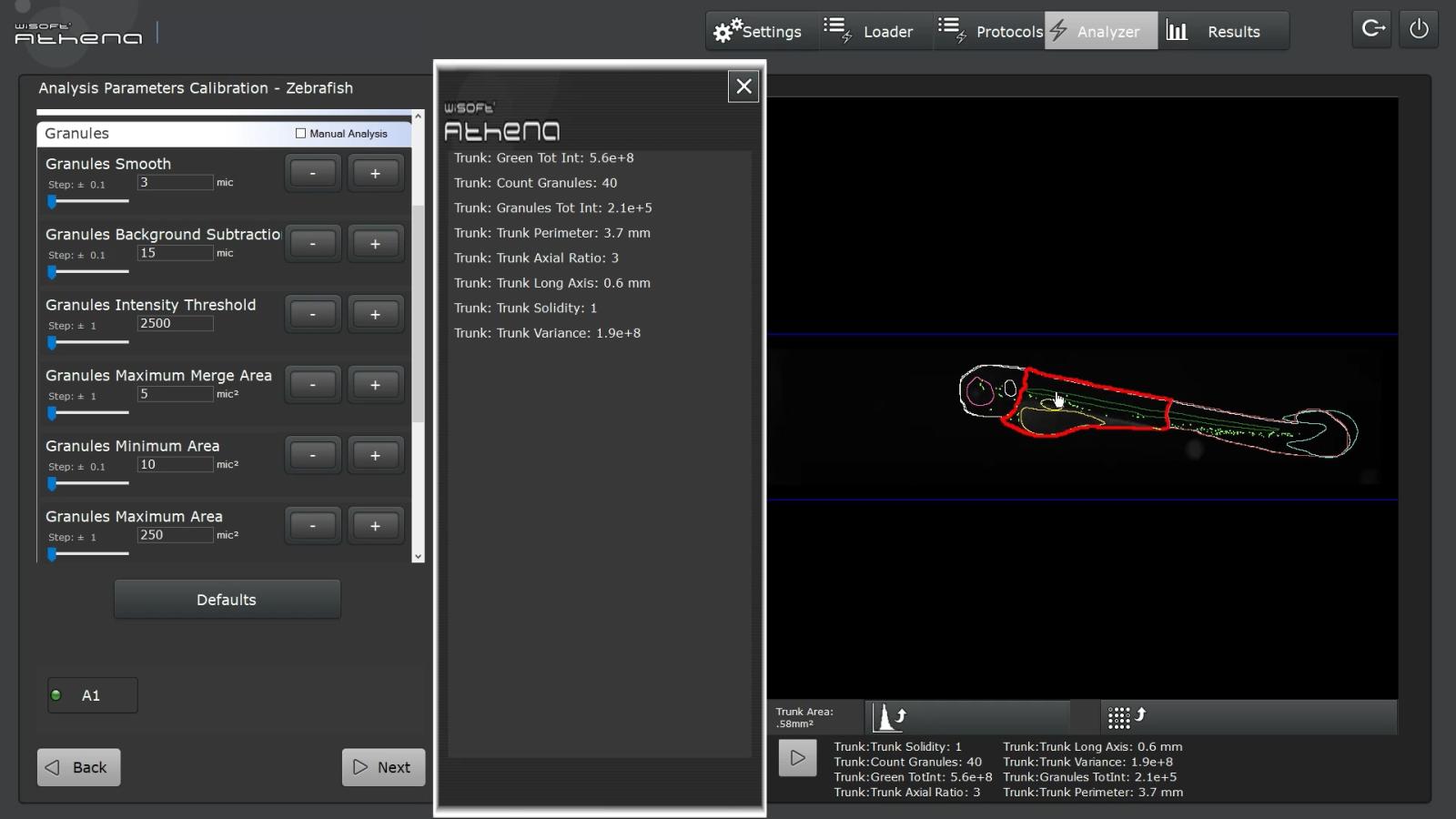
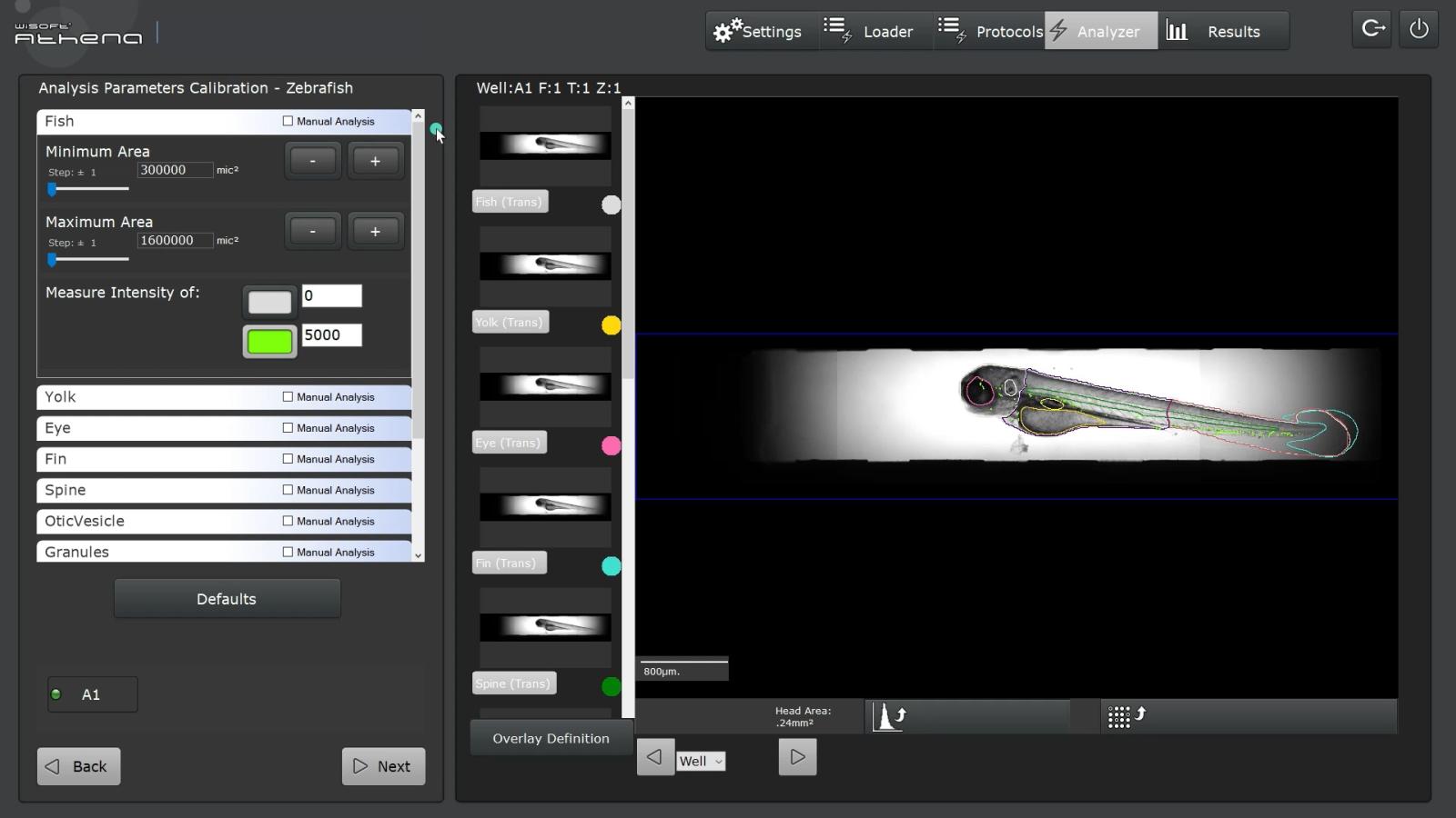
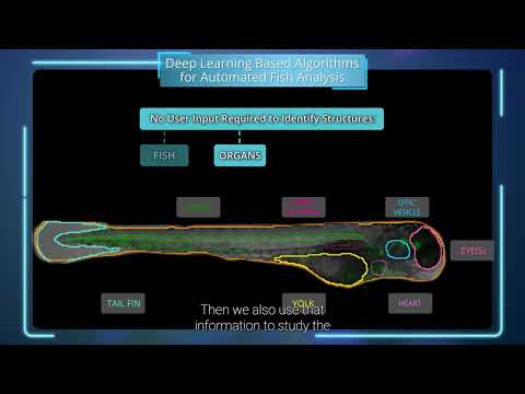
The software permits parameter-free zebrafish analysis using simple bright-field images. It automatically detects zebrafish embryos and larvae up to 5 days old (dpf), extracting the fish contour and much of its internal anatomy: yolk sac, eye, notocord, and more, along with body regions of the head, trunk, and tail.
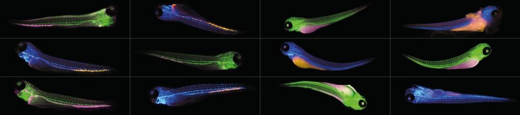
For each of these objects, the software measures the morphology (area, length, and shape) and can detect fluorescence in associated color channels. Both fluorescence intensity and spot/structure detection within specific anatomy are supported.
This new, unique, dedicated analysis software for automated analysis of Zebrafish microscopy images offers simple and quick quantification of fluorescence, measurement of morphological changes & other phenotypic features in Zebrafish larvae in a high throughput format.
Athena Zebrafish is suited for a broad range of researchers and accepts multiple image format types output from nearly all microscope manufacturers.
The software permits parameter-free zebrafish analysis using simple bright-field images. It automatically detects zebrafish embryos and larvae up to 5 days old (dpf), extracting the fish contour and much of its internal anatomy: yolk sac, eye, notocord, and more, along with body regions of the head, trunk, and tail.
For each of these objects, the software measures the morphology (area, length, and shape) and can detect fluorescence in associated color channels. Both fluorescence intensity and spot/structure detection within specific anatomy are supported.
To learn more, visit IDEA Bio-Medical webpage.