Oncology - The importance of oxygen in tumours; accelerating oncology research
Introduction
Understanding tumor oxygen levels is of high importance in preclinical and translational cancer research and treatment, as it plays a critical role in tumor growth, metastasis, and response to therapy. Tumor oxygenation refers to the oxygen availability within the tumor microenvironment, which is a product of oxygen delivery (by perfused blood) on the one hand, and oxygen consumption by tumor cells on the other. By unraveling the intricate relationship between tumor oxygenation and cancer progression, researchers are developing novel therapeutic approaches to target hypoxia, enhance treatment efficacy, and improve patient outcomes.
There are several fields that cancer researchers often directly investigate to help elucidate the links between tumors morphology and oxygen levels while testing novel treatments. At Oxford Optronix, we have supported preclinical and translational research in all the areas listed below. For each area we highlight a selection of publications and provide details of which of our products was involved.
Tumor growth and progression
The oxygen requirements of cancer cells differ from those of normal cells. While normal cells primarily rely on oxygen-dependent metabolism through oxidative phosphorylation, cancer cells often exhibit a metabolic shift known as the Warburg effect. This shift involves increased glucose consumption and reliance on glycolysis, even in the presence of oxygen. The altered metabolism provides cancer cells with the necessary energy and metabolic intermediates to support rapid proliferation and tumor growth. Understanding tumor oxygen levels helps researchers uncover the molecular mechanisms driving the metabolic adaptations in cancer cells and identify potential therapeutic targets to disrupt these processes.
Penjweini R, Roarke B, Alspaugh G, Gevorgyan A, Andreoni, Pasut A, Sackett DL and Knutson JR (2020). Single cell-based fluorescence lifetime imaging of intracellular oxygenation and metabolism. Redox Biol. 34:101549
(OxyLite™ cited)
Andreoni A; Penjweini R; Roarke B; Strub M-P; Sackett DL and Knutson JR (2019). Genetically encoded FRET probes for direct mapping and quantification of intracellular oxygenation level via fluorescence lifetime imaging. Proceedings Volume 10882, Multiphoton Microscopy in the Biomedical Sciences XIX; 108820O
(OxyLite™ cited)
Riemann A, Reime S and Thews O (2017). Tumor acidosis and hypoxia differently modulate the inflammatory program: measurements in vitro and in vivo. Neoplasia. (12):1033- 1042
(HypoxyLab™ cited)
Hypoxia-driven genetic changes
Hypoxia, or low oxygen levels, is a common characteristic of solid tumors. It occurs due to an imbalance between oxygen supply and consumption in rapidly growing tumors. Hypoxia activates a group of transcription factors called hypoxia-inducible factors (HIFs), particularly HIF-1α and HIF-2α. These factors regulate the expression of numerous genes involved in various aspects of cancer progression, including angiogenesis (formation of new blood vessels), cell survival, metabolism, invasion, and metastasis. By understanding tumor oxygen levels and the subsequent activation of HIFs, scientists can gain insights into the molecular mechanisms that promote tumor aggressiveness and develop targeted therapies to counteract hypoxia-driven genetic changes.
Lu N, Zhang M, Lu L, Liu YZ, Liu XD, and Zhang HH (2021). Insulin-Induced Gene 2 Expression Is Associated with Breast Cancer Metastasis. Am J Pathol 191(2), 385-395
(OxyLite™ cited)
Malier M, Gharzeddine K, Laverriere MH, Marsili S, Thomas F, Court M, Decaens T, Roth G, and Millet A (2022). Hypoxia drives dihydropyrimidine dehydrogenase expression in macrophages and confers chemoresistance in colorectal cancer. Cancer Res. 82(7):1436
(HypoxyLab™ cited)
Rauschner M, Lange L, Hüsing T, Reime S, Nolze A, Maschek M, Thews O, and Riemann A (2021). Impact of the acidic environment on gene expression and functional parameters of tumors in vitro and in vivo. J Exp Clin Cancer Res. 2021 Jan 6;40(1):10. doi: 10.1186/s13046-020-01815-4. PMID: 33407762; PMCID: PMC7786478
(HypoxyLab™ cited)
Ritter V, Krautter F, Klein D, Jendrossek V, and Rudner J (2021). Bcl-2/Bcl-xL inhibitor ABT-263 overcomes hypoxia-driven radioresistence and improves radiotherapy. Cell Death Dis 12(7), 694
(GelCount™ cited)
Huang Y et al. (2020). The Impacts of Different Types of Radiation on the CRT and PDL1 Expression in Tumor Cells Under Normoxia and Hypoxia. Frontiers in Oncology, 10
(GelCount™ cited)
Angiogenesis and drug delivery
Tumors require a blood supply to sustain their growth, and they promote the formation of new blood vessels through angiogenesis. Hypoxia within tumors triggers the release of pro-angiogenic factors such as vascular endothelial growth factor (VEGF), which stimulates the development and outgrowth of new blood vessels into the tumor microenvironment. However, the resulting tumor vasculature tends to be structurally abnormal, leaky, and inefficient, leading to inadequate oxygenation. Poor tumor oxygenation not only promotes tumor aggressiveness but also hampers the effective delivery of anticancer drugs. Chemotherapeutic agents and molecularly targeted drugs rely on efficient blood perfusion to reach their target sites within the tumor. Understanding tumor oxygen and the underlying angiogenic processes can help scientists develop strategies to improve blood vessel functionality, enhance drug delivery, and optimize treatment efficacy.
Collet G et al. (2014). Hypoxia-regulated overexpression of soluble VEGFR2 controls angiogenesis and inhibits tumor growth. Mol Cancer Ther 13(1), 165-78
(OxyLite™ cited)
Jordan BF, Sonveaux P, Feron O, Gregoire V, Beghein N, and Gallez B (2003). Nitric oxide-mediated increase in tumor blood flow and oxygenation of tumors implanted in muscles stimulated by electric pulses. Int J Radiat Oncol Biol Phys 55, 1066-73
(OxyLite™ and OxyFlo™ cited)
Kostourou V, Troy H, Murray JF, Cullis ER, Whitley GS, Griffiths JR, and Robinson SP (2004). Overexpression of dimethylarginine dimethylaminohydrolase enhances tumor hypoxia: an insight into the relationship of hypoxia and angiogenesis in vivo. Neoplasia 6, 401-11
(OxyLite™ cited)
Treatment response and resistance
Tumor oxygenation significantly influences the response to cancer therapies such as radiation therapy and chemotherapeutic agents. Radiation therapy relies on the production of reactive oxygen species (ROS) to induce DNA damage and subsequent cell death. However, hypoxic regions within tumors have reduced oxygen-dependent radiosensitivity, making them less susceptible to radiation treatment. Similarly, various chemotherapeutic agents require oxygen to exert their cytotoxic effects. Poor tumor oxygenation can lead to reduced drug efficacy and contribute to treatment resistance. By characterizing tumor oxygen levels, clinicians can predict treatment response and resistance patterns, tailor treatment regimens accordingly, and explore strategies to overcome hypoxia-associated treatment limitations. This may include techniques such as hypoxia-targeted therapies, combining radiation or chemotherapy with oxygen-enhancing interventions, or utilizing alternative treatment modalities specifically designed for hypoxic tumors.
Aiyappa-Maudsley R, Elsalem L, Ibrahim AIM, Pors K, and Martin SG (2022). In vitro radiosensitization of breast cancer with hypoxia-activated prodrugs. J Cell Mol Med. (16):4577-4590
(HypoxyLab™ cited)
Cooper CR, Jones D, Jones GD, Petersson K (2022). FLASH irradiation induces lower levels of DNA damage ex vivo, an effect modulated by oxygen tension, dose, and dose rate. Br J Radiol. 95(1133):20211150
(HypoxyLab™ cited)
Sun H, Ong YH, and Zhu TC (2022). Reactive oxygen species explicit dosimetry (ROSED) for fractionated photofrin mediated photodynamic therapy (PDT). Proc SPIE Int Soc Opt Eng. 11940:1194007
(OxyLite™ cited)
Owen J et al. (2021). Orally administered oxygen nanobubbles enhance tumor response to sonodynamic therapy. Nano Select
(OxyLite™ cited)
Product information
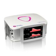
OxyLite – our widely used tissue vitality monitors that measures tumour pO2 in real time via an optical sensor. Sensors are mainly used in xenograft models to either map the oxygen gradient through a tumor or are placed into the tumor to follow the oxygen changes during or after a treatment or therapy (i.e. PDT, oxygen nanobubbles, hypoxic targeted drugs, etc.). The sensors themselves are minimally invasive but rigid enough to be easy to work with. For those needing microvasculature perfusion information, this system also pairs with our OxyFlo counterpart. A unique, combined sensor provides pO2, temperature and a blood perfusion measurement from a single tissue region.
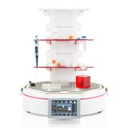
HypoxyLab – is a hypoxic (or physoxic) workstation used by cancer researchers to mimic the oxygen levels seen in vivo in their cellular models. The HypoxyLab allows manipulation within the system via sealed armports that ensure the maintenance of physiological oxygen conditions across a study. Users can control oxygen, carbon dioxide, humidity and temperature on a front facing touchscreen. Uniquely, the HypoxyLab ensures that oxygen levels are comparable and reproducible week to week and between laboratories in different geographical locations by maintaining and controlling internal oxygen conditions in absolute units of partial pressure (mmHg). (For further reading: The Reproducibility Issue within Hypoxia Chambers and a Simple Solution to Fix it).
GelCount – our popular colony counting platform used by cancer biologists employing the colony, spheroid or organoid formation assay. The GelCount is commonly used to quantify the cytotoxicity of hypoxia, drugs, and other treatment regimes, often in combination, in tissue culture models of cancer growth. It is a powerful labour-saving device capable of unlocking substantial throughput improvements in laboratories undertaking these assays at scale.


