MOM® — Open-Design, Multi-Photon Microscope
MOM® Movable Objective Microscope
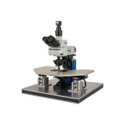
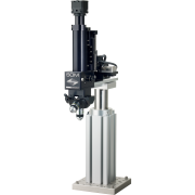
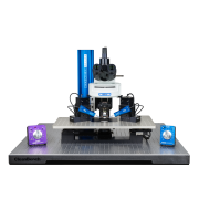
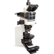
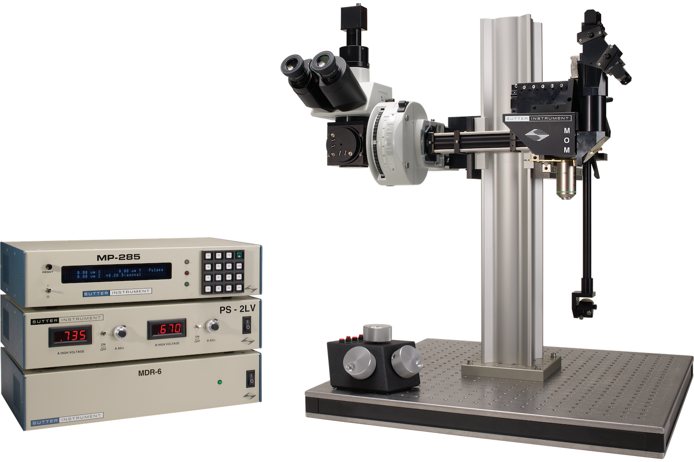
The MOM® (Movable Objective Microscope) from Sutter Instrument is a fully customizable two- and three-photon imaging platform that has been the reference open-design multiphoton microscope in neuroscience for over two decades. Its central innovation — a motorized objective that translates 22 mm in X, Y and Z and rotates about the optical axis — keeps the specimen stationary during imaging. This removes the mechanical instability introduced by moving stages and permits imaging of non-horizontal tissue surfaces and volumes without compromising optical alignment.
Opto-Mechanical Design
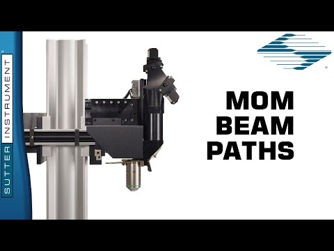
The MOM is built around two independent optical paths sharing the same motorized objective stage. The wide-field side provides epifluorescence via an Olympus vertical illuminator and Sutter Xenon arc lamp with a camera port for standard fluorescence imaging. The two-photon/three-photon side delivers the pulsed laser beam from the table through galvanometric or resonant scanners, the scan and tube lenses, and into the back aperture of the objective. Two coupled steering mirrors maintain efficient beam delivery regardless of the objective's position or rotation angle. The main body retracts on a rail for unobstructed access to the preparation before imaging.
Scanning Systems
The MOM supports the full range of current scanning technologies: Cambridge Technology 3 mm or 6 mm XY galvo scanners, resonant-galvo, and resonant-galvo-galvo configurations including the Vidrio RMR scanner. New scanning hardware can be retrofitted into existing MOM frames with minimal changes, protecting the long-term value of the platform.
Imaging Software
The MOM is compatible with multiple software packages. Sutter's own MScan 3.0 (part of the MOM Computer System, MCS) is Windows 10 compatible. The MOM is also fully compatible with Vidrio ScanImage Premium, ScanImage Basic, and ScanImage freeware. Data acquisition hardware is available for NI PXI FPGA, Vidrio vDAQ, and NI PC-based multifunction I/O platforms. Conoptics Pockels cells are used for laser intensity control.
Applications
- Two-photon and three-photon deep-tissue imaging — the moving-objective design eliminates stage-induced drift, making the MOM the preferred platform for long-duration in vivo calcium imaging, voltage imaging and optogenetics in head-fixed animals. Objective rotation allows imaging of tilted or curved tissue surfaces inaccessible with a conventional fixed-objective upright.
- In vivo and in vitro combined experiments — the specimen remains stationary at all times, so the same preparation can be imaged at multiple depths and positions without repositioning the animal or the recording electrodes.
- Three-photon imaging — the MOM has an established record in 3P imaging at 1300 nm and 1700 nm, including pioneering work in Chris Xu's group at Cornell and ongoing use at the Allen Institute of Brain Science. Sutter can configure 3P-MOM systems with optics and detectors optimised for longer-wavelength excitation.
- Objective travel (X, Y, Z): 22 mm on all three axes
- Objective rotation: 0–180° about the optical axis
- Resolution — MPC-200 controller: 0.0625 µm/step
- Resolution — MP-285 controller (Low/High): 0.2 µm/step / 0.04 µm/step
- Maximum speed — MPC-200: 5.0 mm/sec
- Maximum speed — MP-285: 2.9 mm/sec
- Long-term stability: 1–2 µm/hour
- Drive mechanism: Precision worm gear capstan drive
- Communication (MPC-200): USB
- Communication (MP-285): RS-232 Serial
- Electrical: 115/230 V, 50/60 Hz
- MOM-BASIC MOM basic system — includes moving objective microscope, 2-channel detector with PMTs, preamplifiers and PS-2 power supply, XY scanners with drive electronics, wide-field fluorescence unit with vertical illuminator and 300 W Xenon arc lamp