SOM® — Son of MOM®, Simple Moving Microscope
SOM® — Son of MOM® Simple Moving Microscope
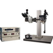
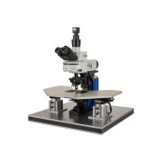
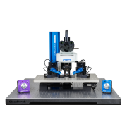
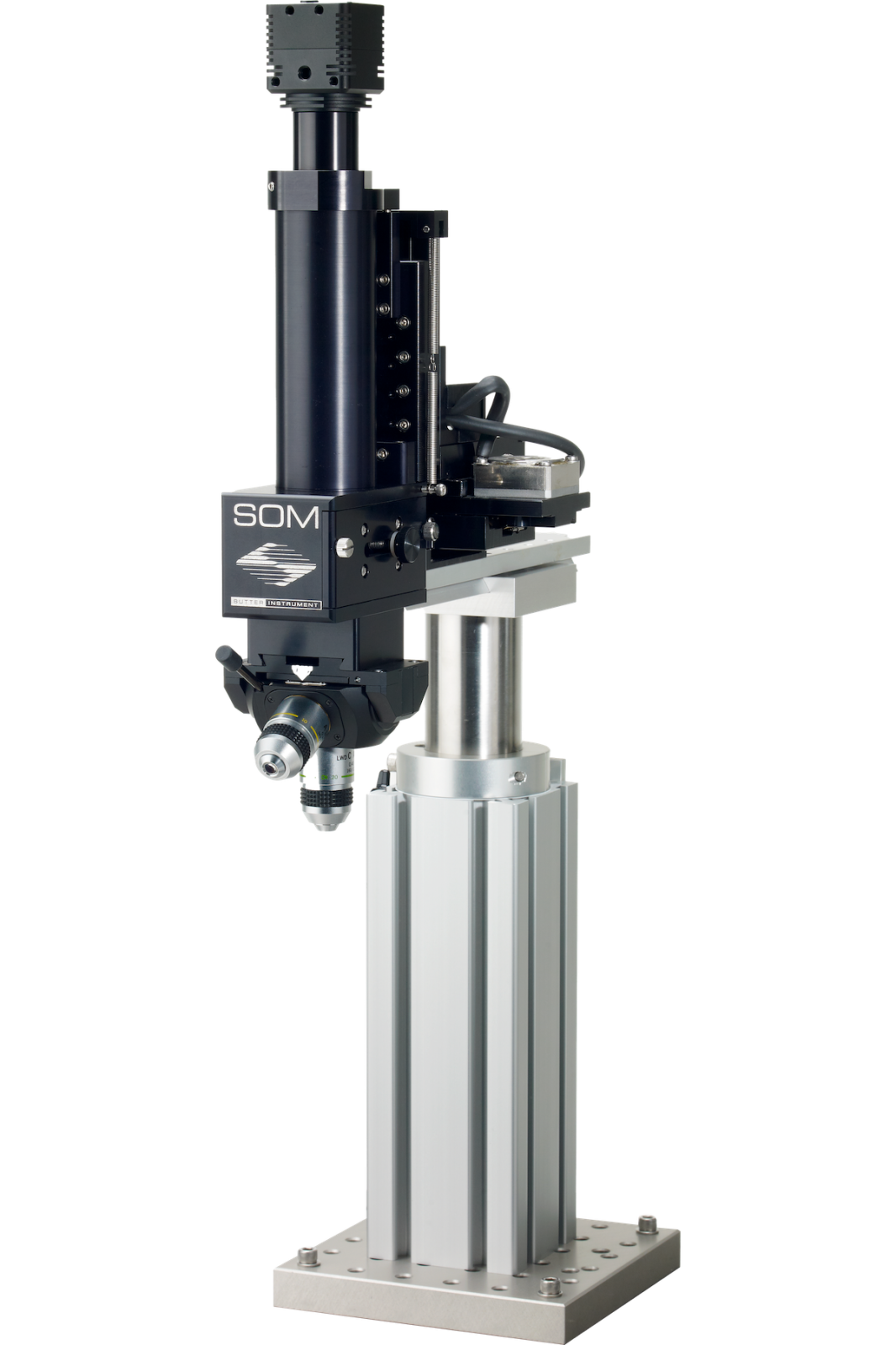
The SOM® (Son of MOM®) from Sutter Instrument is a compact, robotically-positioned microscope that adapts the moving-objective concept of the MOM® to single-unit and patch-clamp electrophysiology. Rather than moving a stage or the sample, the entire microscope body is carried by an MP-285 or MPC-385 motorized micromanipulator. X, Y and Z movements of the manipulator position the objective over the sample and bring it to focus — eliminating large translators, moving stages and the mechanical noise they couple into recording electrodes.
Robot-Driven Positioning
Because the SOM rides on the same manipulator platform as Sutter's recording electrode positioners, the free Multi-Link™ software can coordinate microscope and manipulator positions simultaneously. In a slice recording session, once you have searched the slice and found a target neuron, Multi-Link retrieves the recording and stimulation pipettes directly to that field of view without the experimenter having to search again. If you need to stimulate a region outside the current field, the software locks the recording pipette position, moves the objective and stimulating pipette(s) to the new location, then allows you to return.
Imaging and Illumination
The SOM uses transmitted IR LED illumination combined with an IR-capable CCD camera — sufficient for the vast majority of in vitro electrophysiology tasks. A built-in two-position filter cube accommodates fluorescent cell identification or photostimulation filter sets. An optional OCC condenser that translates with the microscope in X and Y maintains consistent illumination during repositioning. The fluorescence excitation port has C-mount threading and standard cage-component mounting holes, allowing custom light sources to be coupled to the excitation path.
Applications
- Combined in vivo / in vitro electrophysiology — the robotically-moved microscope eliminates the physical footprint of large stages beneath the objective, making in vivo access to head-fixed animals on the same rig used for brain-slice work straightforward. No physical reconfiguration of the rig is needed between modalities.
- Whole-cell patch-clamp recordings — movement is smooth, drift-free and reproducible to 0.0625 µm/step, matching the quality of a precision micromanipulator. Mechanical coupling between the microscope and the recording electrode is eliminated because both are driven by independent motors on the same rigid base.
- Space-constrained laboratories — removing the translators and stages from below the objective substantially reduces the overall rig footprint, making the SOM the preferred microscope platform when every centimetre of optical table space matters.
- Controller options: MP-285 or MPC-385 motorized micromanipulator
- Resolution — MPC-200/MPC-385: 0.0625 µm/step
- Resolution — MP-285 (Low/High): 0.2 µm/step / 0.04 µm/step
- Maximum speed — MPC-200/MPC-385: 5.0 mm/sec
- Maximum speed — MP-285: 2.9 mm/sec
- Objective thread: RMS (standard); contact Sutter for other options
- Filter cube positions: 2
- Electrical: 115/230 V, 50/60 Hz
- SOM-MP285 Son of MOM® microscope with MP-285 controller
- SOM-MPC385 Son of MOM® microscope with MPC-385 controller