GelCount™ - automated cell colony counter
Mammalian cell colony counter for organoid and spheroid models, organoid growth, and organoid cell culture.
Second-generation imaging and analysis platform for 3D cell colonies, spheroids and organoids in multi-well plates and Petri dishes
- High-resolution whole well or whole dish view, no z-stacking
- Substantial throughput and objectivity benefits over manual microscope counting
- Generate object counts AND diameter statistics per well or dish
- Direct exportation of output data to Excel®
- Unlimited software installations for image processing on other workstations
The benchmark colony counter for spheroid and organoid detection and analysis
The colony formation assay and colony counting is universally recognized as the gold standard method for measuring the effects of radiation, chemotherapeutic drugs and other agents on cancer cell viability and testing.
Meanwhile, the growth of multi-cellular 3-dimensional organoids, derived from primary cell types that mimic the structure and function of human tissues and organs, has revolutionized the study of disease and therapeutic responses in vitro.
However, manual detection and analysis of the resulting colonies, spheroids or organoids, typically under a microscope, is a thankless, laborious and rate-limiting task in which consistency and objectivity are difficult to achieve.
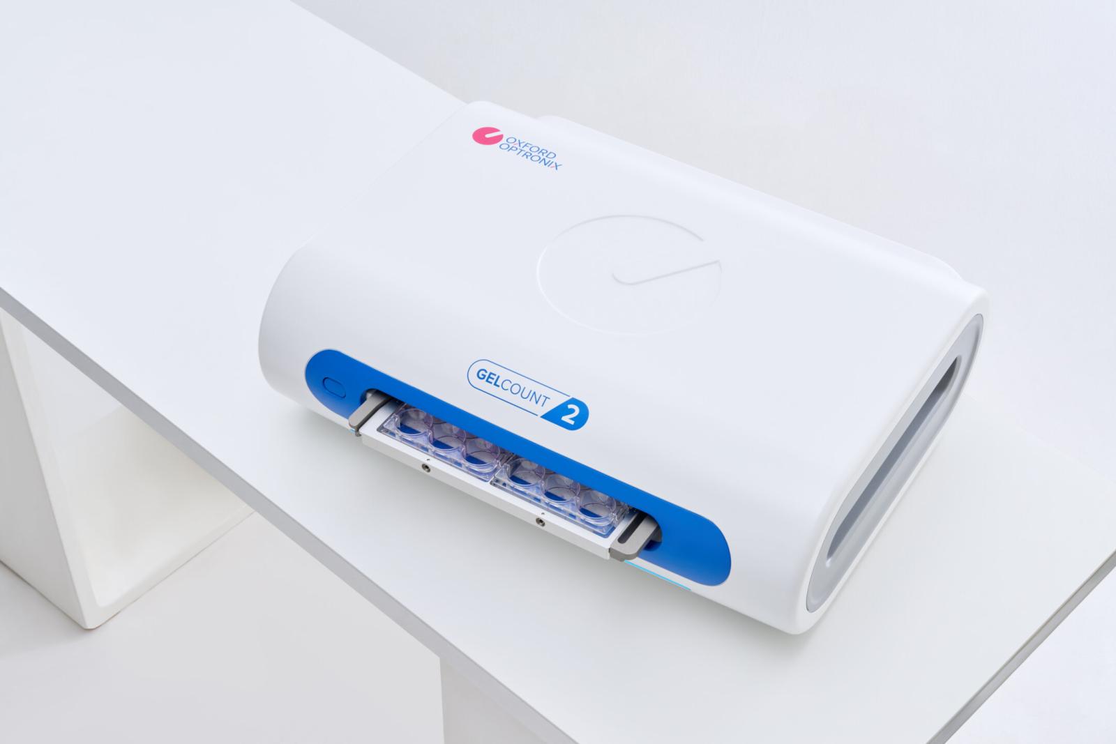
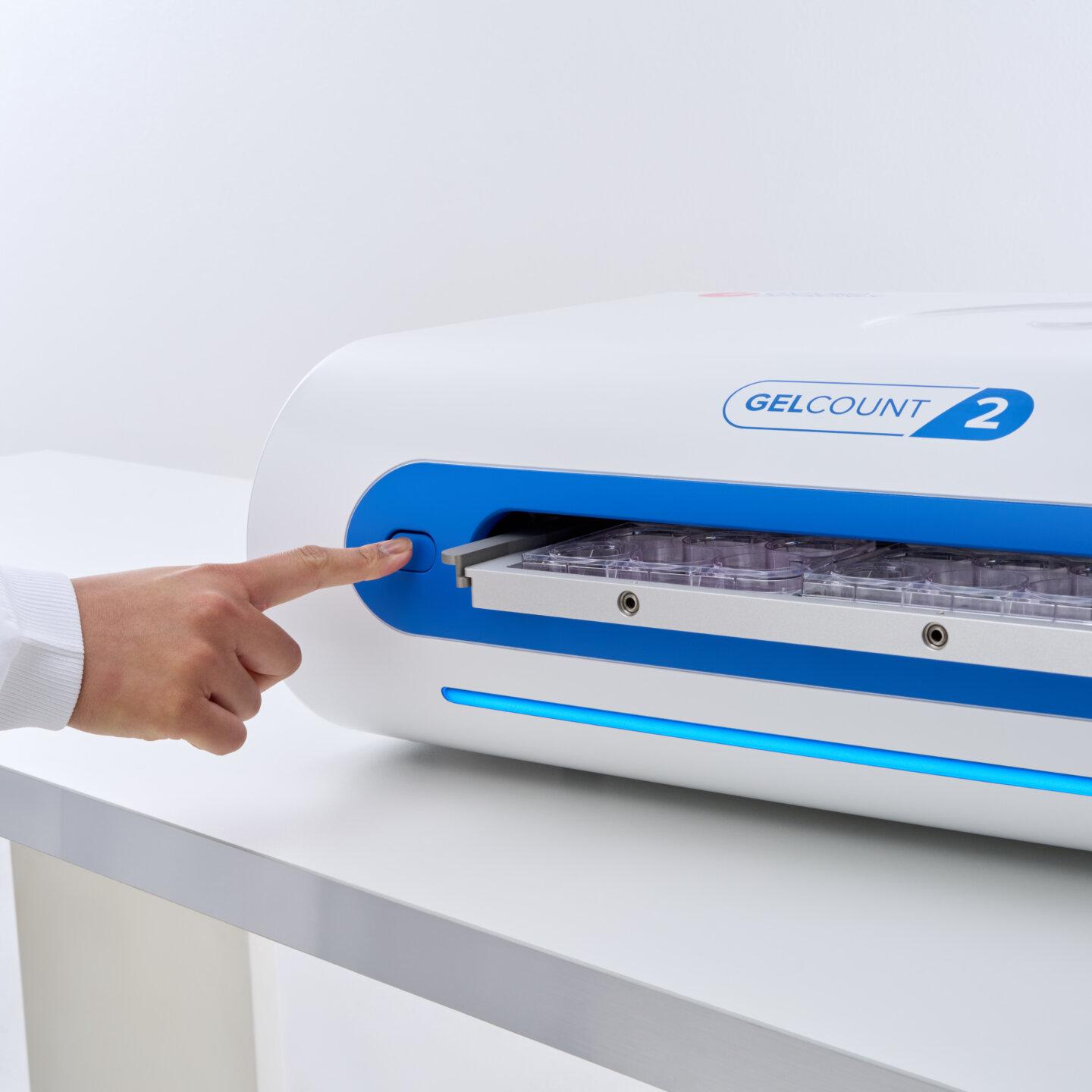
GelCount™ is an intuitive, PC software-operated imager that automates the detection, counting and analysis of mammalian cell colonies, organoids or spheroids in multi-well plates and Petri dishes.
With over 500 scientific citations to its name GelCount™ has become a go-to analysis solution for biologists working with organoid models or employing the colony formation assay.
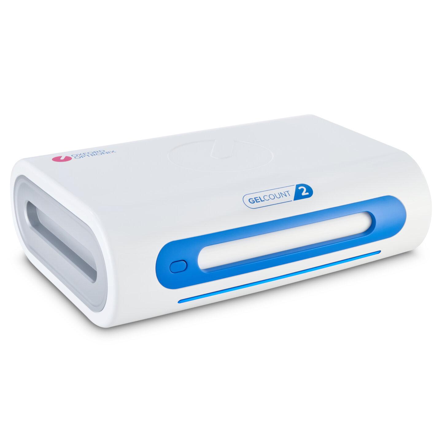
GelCount™ 'Generation 2' introduces a revised design, reduced imaging times, and a more intuitive and powerful software experience, building on the legacy of the original.
An all-in-one imaging and analysis platform for mammalian colonies, spheroids, 3d culture and organoids
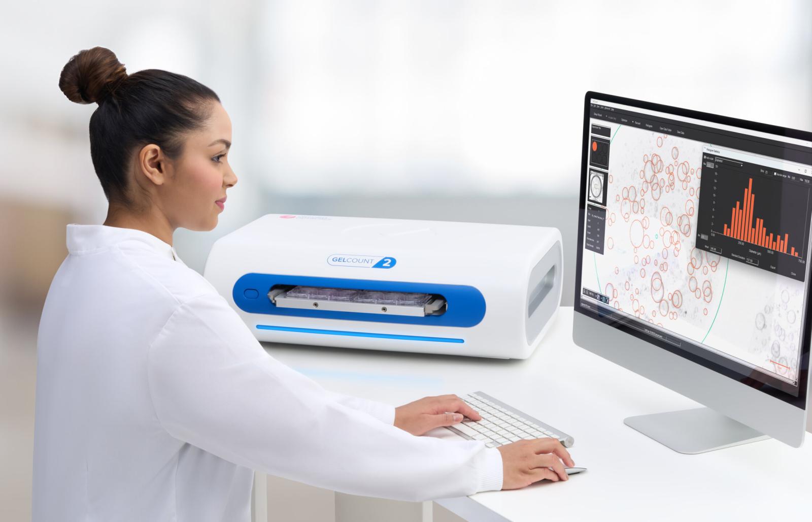
GelCount is an integrated solution for the imaging, detection and characterization of adherent or 3-dimensional colonies, spheroids or organoids on a single integrated hardware and accompanying PC software platform. Culture plates are imaged, images transferred to a PC, images processed and characterized, and the data collated/exported from within a single, integrated user interface.
The industry benchmark
With a track record of over 500 peer-reviewed citations and counting, and a worldwide user-base, the GelCount has become the solution of choice for biologists employing various guises of the colony, spheroid, or organoid formation assay.
Objective, unbiased output
With GelCount the user goes from colony sample to colony counts, colony size distribution and a host of additional statistical data at the click of a button.
GelCount thereby not only dramatically enhances throughput but its inherent 'machine' objectivity and consistency eliminates human error due to subjective interpretation, bias or plain fatigue – a particularly acute problem when manually processing spheroids and organoids under a microscope.
High-resolution on-screen view
Entire wells or Petri dishes can be conveniently viewed on-screen at high resolution. Three dimensional spheroid or organoid cultures are imaged across a media depth of up to approximately 5 mm (subject to contrast conditions) without the use of z-stacking.
Performance of GelCount
Using single pass, high depth-of-field line imaging, combined with a single-axis motorized sample-carrier mechanism, GelCount provides unsurpassed object detection performance including resolution of overlapping objects and differentiation of real colonies from debris or other artefacts.
A typical experiment consisting of four 6-well plates containing spheroids or organoids in 3D culture can be imaged and processed in less than 10 minutes, including user-definable data and image exportation.
The diameter advantage
Processing with the GelCount not only generates a numerical count or colonies, spheroids or organoids but crucially also yields object diameter information in the form of a mean per well/dish, a histogram distribution, or even on a per-object basis if required.
The ability to quantitatively measure the effects of a therapeutic regime not only on absolute cell aggregate numbers but also on their size provides hitherto unavailable insight relating to cellular growth dynamics.
Versatility of GelCount
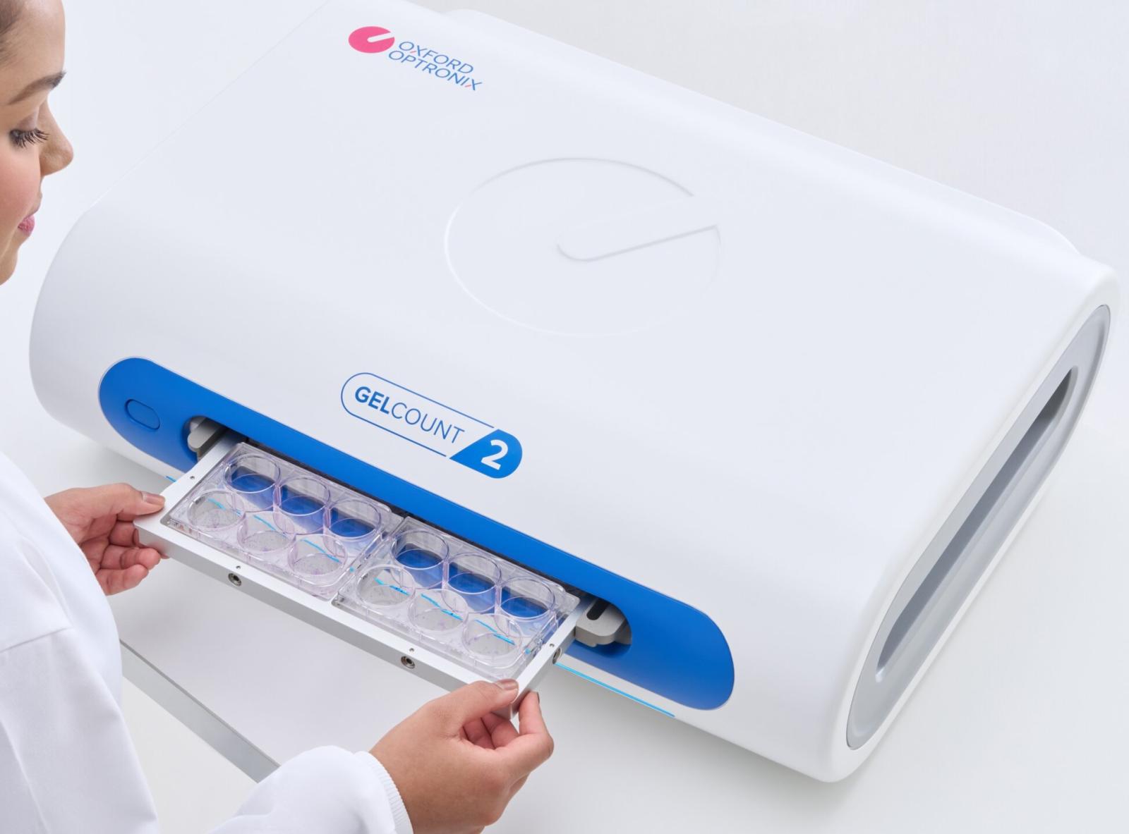
GelCount is suitable for imaging and processing both adherent (typically stained) cell colonies, and non-adherent (typically unstained) cell aggregates in 3D suspension or semi-solid media, such as spheroids and organoids. Up to 4 multi-well plates or up to 24 Petri dishes can be imaged simultaneously.
Workflow and throughput optimisation
Not only does GelCount represent a single integrated hardware and software platform for imaging and processing of cell growth assays, but the software can also be installed on unlimited other workstations. Images generated by GelCount can therefore be stored, transferred to and processed ‘offline’ at the user’s convenience on any other workstation, without tying up the imager for other users.
Flexible data output options
Colony / spheroid counts, diameter statistics and other numerical data are automatically exported to Excel®, while colony images can be saved in a raw format for subsequent offline processing or in a generic image format for printing, presentations, etc.
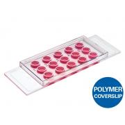
- PETRY-TRAY 4-place tray for multi-well plates
- PETRI_TRAY_100 4-place tray for 100 mm Petri dishes
- PETRY TRAY 50 12-place tray for 50 mm Petri dishes
- PETRI_TRAY_35 24-place tray for 35 mm Petri dishes