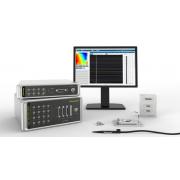
Electrophysiology: Cardiac Electrophysiology – Mapping Systems
Imagine if a scientist could pinpoint a certain area of a diseased heart, which may be responsible for the cause. This would lead to better treatment selection and a higher recovery rate.
What if pharmaceutical companies could screen any drug that may cause harm to the heart? They would provide lifesaving therapies more efficiently, with better treatment and saved resources.
This is possible with mapping technology.
In the past, it was a mystery to measure and examine the action potential from the endocardial or epicardial surface of the heart. Despite the presence of bipolar and unipolar recording, mapping systems provide the most accurate way to examine the physiological activity of the heart at the tissue or organ level (3).
Let’s dive into both electrical and optical mapping below.
What is Optical Mapping?
Cardiac optical mapping has been recognized as a vital technique to examine and investigate the mechanisms of cardiac arrhythmias. This technology uses fluorescence-based imaging to investigate the electrical properties of multicellular cardiac preparations. It is designed to measure important physiological parameters, such as action potential or calcium transient, at high spatiotemporal resolution
Moreover, this technique explores new horizons in terms of biological samples, which includes (but is not limited to):
-
Isolated hearts
-
Intact cardiac tissue preparations
-
Muscle slice
-
Monolayers of pluripotent stem-cell-derived or neonatal cardiac myocytes
But how does it work?
First, the isolated heart is introduced to a non-beating state. Voltage-sensitive dyes keep track of the fluctuations in the action potential difference across the cellular membrane of cardiac myocytes. Meanwhile, calcium-sensitive dye images the spread of electrical waves in the heart (calcium transient).
Optical mapping has great potential to generate high spatiotemporal resolution information on electrophysiological properties. This makes it a popular technique to interrogate the basic mechanisms implying the electrical stability of heart diseases (4,5).
For cardiology, an optical mapping system dissects the basic mechanism which governs the electrical stability of the heart to control and examine different cardiac diseases. With the help of high-speed cameras and fluorescence microscopy, the fluorescence fluctuations are recorded on the epicardial or endocardial surface.
Benefits of Optical Mapping
-
Efficient Quality Data Collection
Optical mapping is a high-resolution, less invasive method to study electrical cardiac activity. It is able to generate tons of high quality data in a short period of time.
-
Measures Simultaneous Multiple Electrophysiological Parameters
Optical mapping is a simple way for the imaging process of electrophysiological activity comprising the whole outer of Langendorff-perfused hearts. Currently, optical mapping is the only known method to examine a number of primary electrophysiological parameters. (6)
- Wide Range of Detectable Sensor Dye
The application range of fluorescent dyes has been extended to magnesium, sodium, potassium, pH, nitric oxide, and redox state or oxygen levels. Optical mapping is integrated into a powerful system that allows the acquisition of a more comprehensive dataset. This is extremely important for future mechanistic drug design.
What is Electrical Mapping?
Electrical mapping technology uses physical electrodes to measure and study the electrical properties of multicellular cardiac preparations. This system is specially designed to record electrical activity and quickly provides conductive data.
Typically, with the help of a multiple electrode array (MEA) as well as sophisticated depolarization/repolarization calculations, this system is capable of detecting the abnormalities in different elements like sodium, potassium and calcium currents. The development of measuring electrodes for a variety of cells/tissues will certainly change our way of investigating. Researchers and analysts will have a better understanding of cardiac arrhythmias, which includes atrial fibrillation and ventricular fibrillation.
Multifaceted Benefits:
Electrical mapping technology accurately records depolarization and repolarization of the cardiac action potential at various regions of heart from in vivo and ex vivo preparation. In addition, it can monitor cardiac pacemaker activity as well as electrical conduction/velocity.
Cardiac electrical mapping provides information such as:
- Initiation Sites
- Frequency of Action Potentials
Direction and Velocity of Conduction
Electrical Conduction Dispersion
Repolarization Dispersion
QT Interval Dispersion for Both In Vitro and In Vivo Experimental Sampling
Drug Screening
If you are looking for a quick drug screening for cardiac action potential and electrical conduction events, electrical mapping is an excellent method to get the job done. It gives a quick and full-capacity analysis on all electrical waves at probed regions.
On the other hand, optical mapping is a powerful tool to explore mechanistic drug design and it has wider applicability in terms of discovering new drugs, especially in the field of cardiac development. It extracts more authentic and detailed data with the help of fluorescent dyes.
The best thing is these two mapping system can simply work together to harness the full potential of mapping technology. While electrical mapping can be used to monitor electrophysiological parameters, optical mapping can do its job more efficiently and precisely.
Moreover, the latest research and medical experts have defined optical mapping as a reliable tool to screen drug toxicity as it has a sound capability to provide direct electrophysiological insights on new drugs (7,8,9).
Final Words
This article provides a short overview of both optical and electrical mapping systems and their applicability to cardiac examination and analysis. These mapping technologies are gaining more attention in the cardiology field. They can acquire valuable pre-clinical data fast, allowing researchers to explore deeper insights to treat heart diseases. In the long term, they will play a vital role in drug development as the timeframe for drug toxicity screening will be greatly reduced. This would undoubtedly save pharmaceutical companies millions of dollars before entering clinical trials, and could potentially save millions of lives in the process.
Reference
- Heart Disease and Stroke Statistics—2015 Update A Report From the American Heart Association
- Sudden cardiac death: epidemiology and risk factors
- Ventricular resection guided by epicardial and endocardial mapping for treatment of recurrent ventricular tachycardia
- Optical imaging of voltage and calcium in cardiac cells & tissues
- Three‐dimensional integrated functional, structural, and computational mapping to define the structural “fingerprints” of heart‐specific atrial fibrillation drivers in human heart ex vivo
- Low-cost optical mapping systems for panoramic imaging of complex arrhythmias and drug-action in translational heart models
- Optical electrophysiology for probing function and pharmacology of voltage-gated ion channels
- Bringing the light to high throughput screening: use of optogenetic tools for the development of recombinant cellular assays
- OptoDyCE as an automated system for high-throughput all-optical dynamic cardiac electrophysiology