19-20.03.2024, Poznań, Poland
A Charles River-Hosted JAX® Webinar
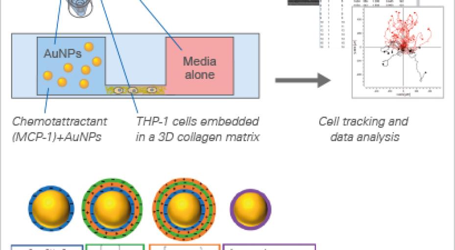
ibidi Blog | January 31, 2024 | Abhishek Derle, ibidi GmbH
Drug Screening
Nine Questions Every Organiza
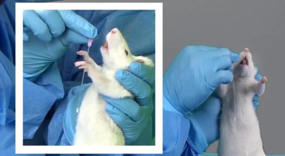
Everything you need to know about using flexible feeding tubes for rodents
Fiber Photometry System
Good and reliable surgical instruments are important tools for optimal...
by Justin Croft, September 2023
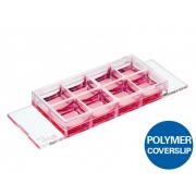
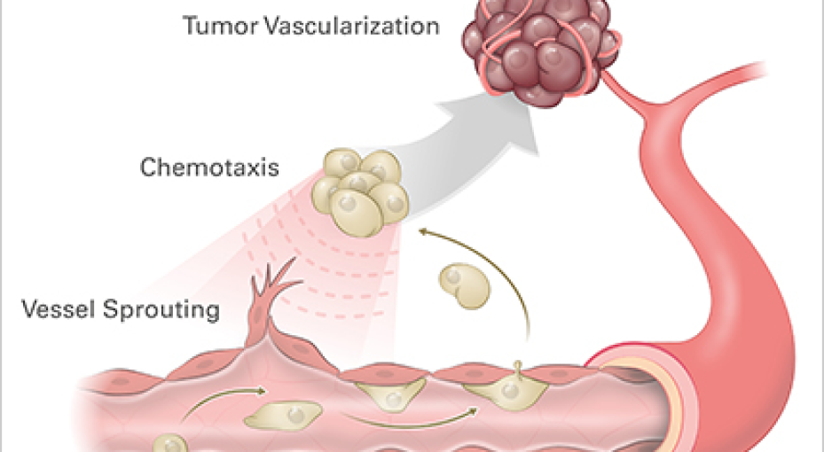
Endothelial cells (ECs) are specialized cells that line the inner surface of...